This is a fast fact based on this post. Full reference available there.
This is a fast fact based on this post. Full reference available there.

FAST FACT – This is based on the Burns: The Practical Stuff video and post.
Full reference available in full post.

FAST FACT – This is based on the Burns: The Practical Stuff video and post.
Full reference available in full post.
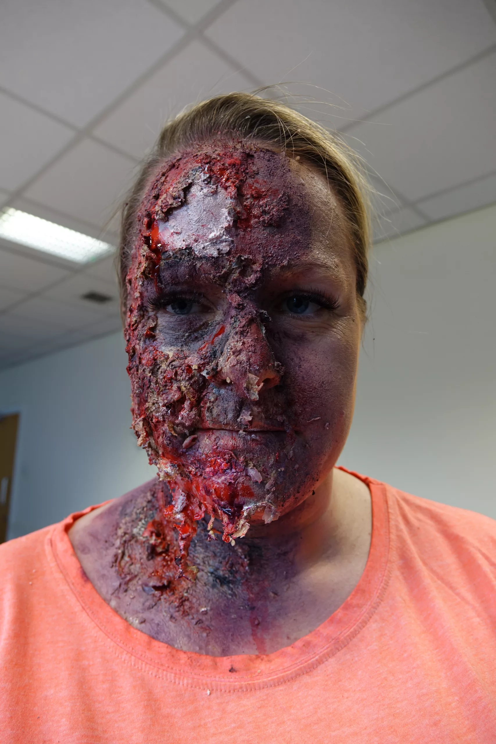
Content warning: This video may contain sensitive content.
This video demonstrates the assessment and management of burns, using equipment that is recommended within the locality at the time of production. Remember to check your local, current guidelines before putting anything into practice. Produced by HCPs and students.
This video demonstrates use of burn gel wraps, which may no longer be used in your ambulance Trust. Please check your local guidelines regarding this. According to the British Burn Association:
Burn gel wraps may be used to provide analgesia, but only after adequate cooling has
Varley et al (2014)
occurred as they do not actively remove heat from the wound
Remember to follow good IPC in real life.
The transcript below has been slightly edited, with titles added, to make it easier to read.
Further recommended reading: The British Burn Association First Aid Clinical Practice Guidelines
You must read the full disclaimer at www.article999.co.uk/disclaimer and check your local guidelines before putting into practice any of our content.
BURNS: The practical stuff
This video contains content some might find sensitive.
With all assessments and interventions, use an aseptic technique, especially because ‘burns are prone to infection’
Purcell, 2003: 217

The patient’s airway might worsen. Here’s why:
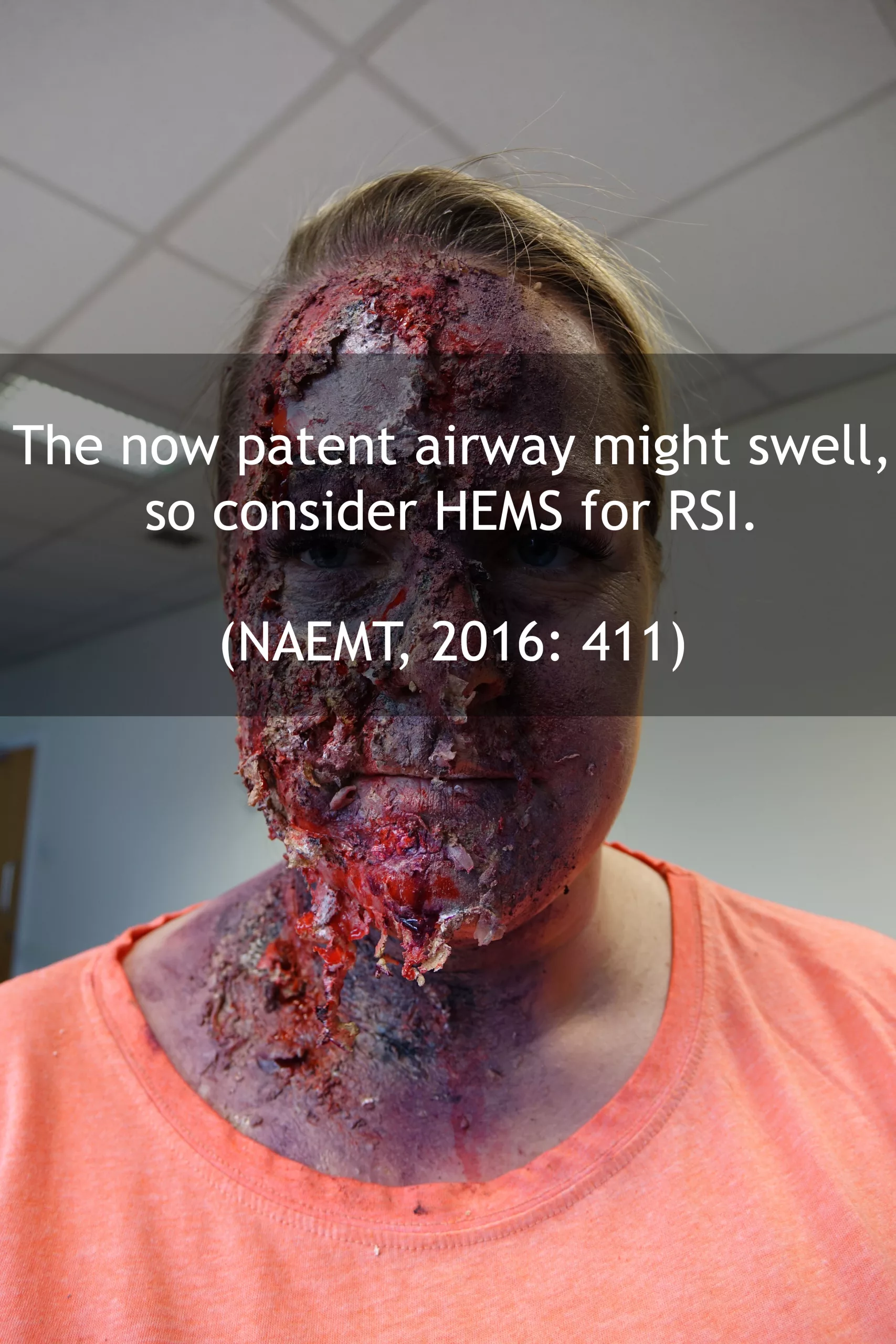
‘A burned patient may have a patent airway on the initial evaluation. […] In the time that follows, the face, as well as the airway, will likely swell.’
NAEMT, 2016: 411
… So, consider HEMS for RSI.
The patient’s breathing might worsen. Here’s why:

Is there a chest wall burn?
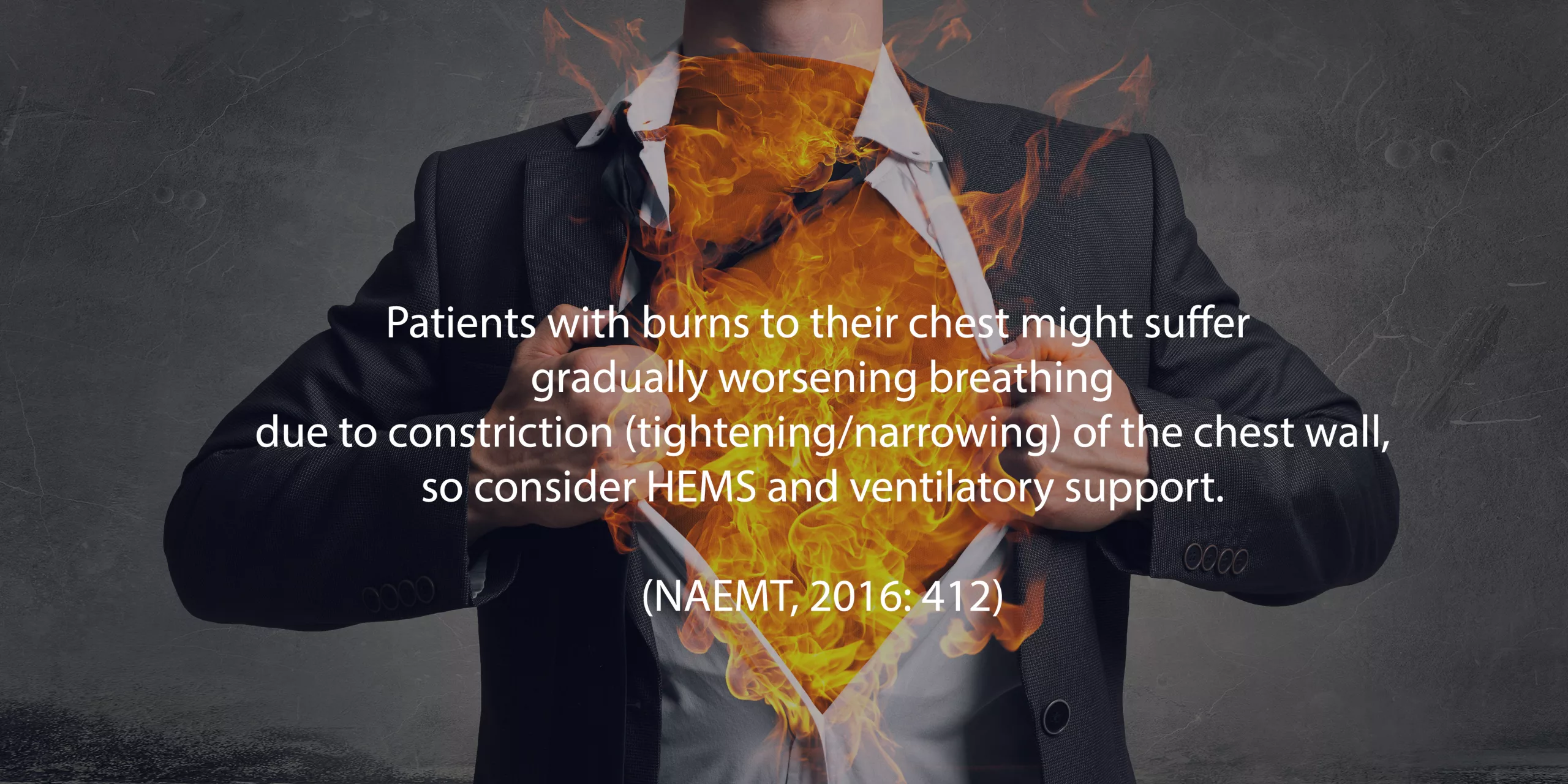
‘Burns constrict the chest wall similarly to having several leather belts progressively tightening around the patient’s chest. As time progresses, the patient cannot move the chest wall to breathe.’
NAEMT, 2016: 412
So, consider HEMS and ventilatory support where needed.
Other considerations:
Inhaled toxins ‘can produce asphyxiation’
NAEMT, 2016: 412
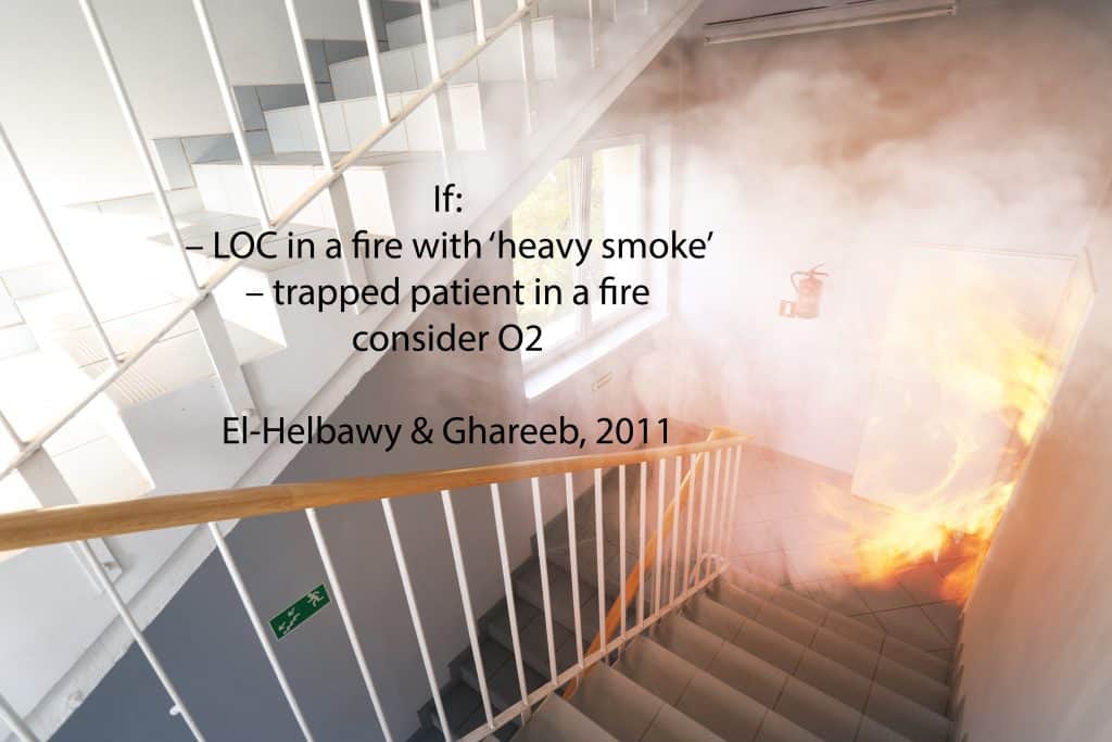
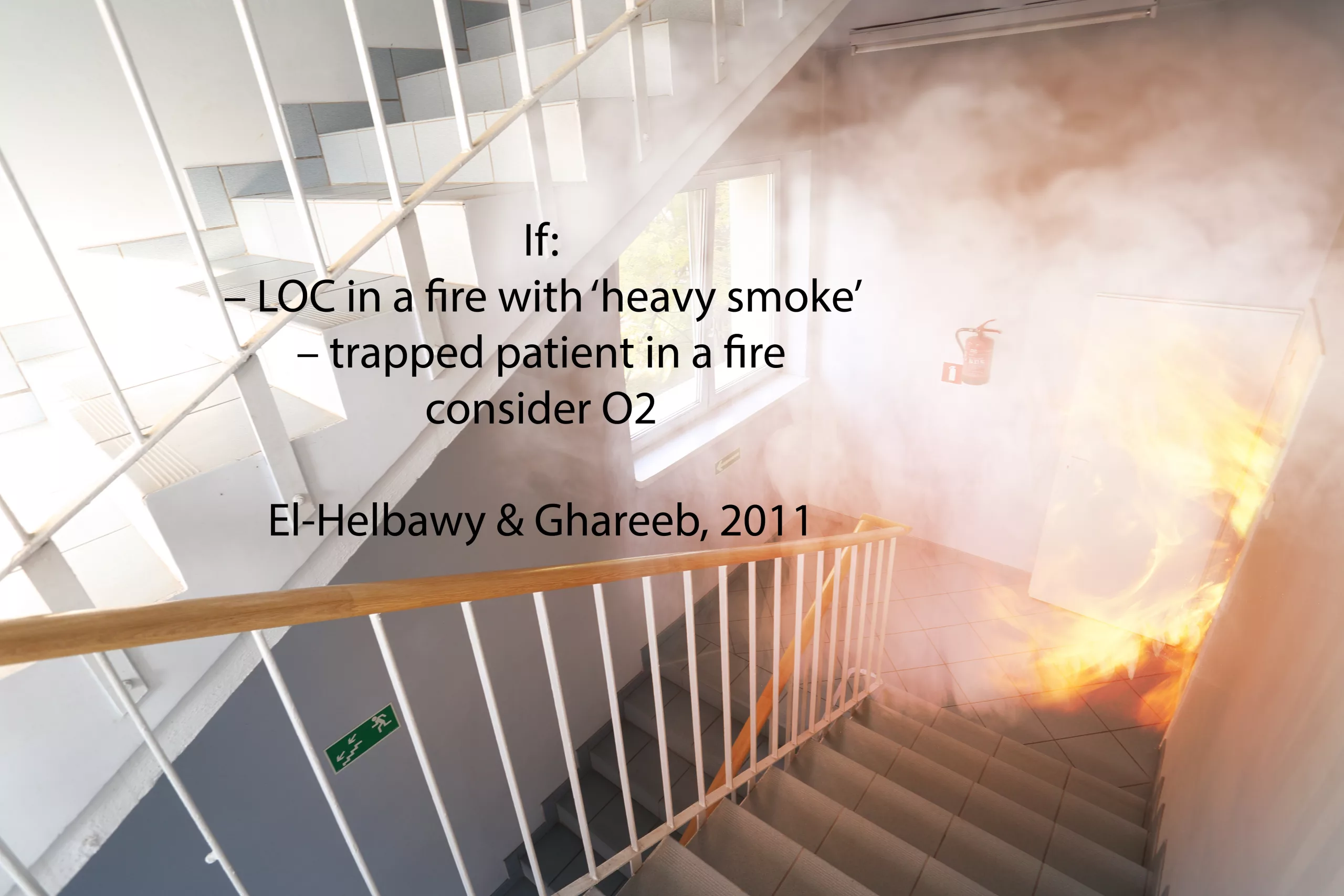
If:
El-Helbawy & Ghareeb, 2011
– LOC in a fire with ‘heavy smoke’
– trapped patient in a fire
consider O2

Swelling might get worse. Here’s what to do about it:
‘Distal limb perfusion may be critically reduced’; ‘Burned extremities should be elevated during transport to reduce the degree of swelling in the affected limb’
NAEMT, 2016: 412
A complication of swelling:
Fluid loss occurs from swelling and evaporation
NAEMT, 2016: 416
Giving Fluids – Pros and Cons
Consider IV fluids, especially if the burn is >10% of the body (AACE, 2016: 265-266). Keep watching for info on how to determine this.
Give fluids with caution. Too much -> ‘cardiac failure, […] infectious complications, acute respiratory distress syndrome, and even death’.
Too little -> ‘hypovolemic shock, organ failure’
(NICE, 2016)
Judicious fluid management of children with severe burn injury can improve the respiratory outcome measures of these children
Duran and Sheridan, 2016
Cannulating – Essentials
When cannulating, ‘placement through the burn is appropriate [only] if no alternative sites are available’
NAEMT, 2016: 412
What about heat?
Patients with burns are not able to maintain their own body heat
NAEMT, 2016: 413
…So, give blankets

ECGs
ECGs are required for electrical burns (AACE, 2016: 265) but consider them for all burns as ‘cardiac dysrhythmias’ result from the release of ‘muscle potassium’ (NAEMT, 2016: 418) and studies have shown disturbances in the cardiac functions of in-hospital burn patients (Jeschke et al, 2008).
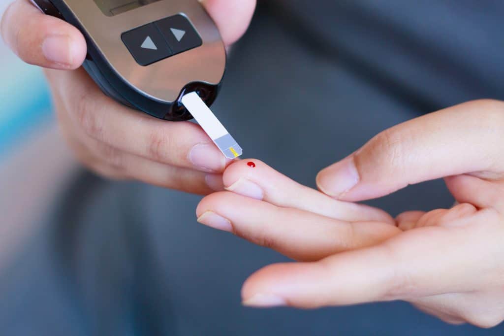
Do your BMs
Hyperglycemia may occur in burns patients and has a higher risk of ‘morbidity and mortality in critically ill patients’ (Wolfe et al, 1979; Mecott et al, 2010).
Pain management in children
In children, ‘a multi-modal approach to pain and sedation can improve the neurological status of severely burned children’
Duran and Sheridan, 2016
Cooling the burn:
Cool with a wet compress
Purcell, 2003: 217
‘Cooling gels such as Burnshield are often used by paramedics. These are useful in cooling the burn and relieving pain in the initial stages.’ *
Be aware of the risk of ‘heat loss’
Hudspith and Rayatt, 2004
*This guidance is changing. Check your local guidance first, and consider the use of cling-film after running water.
Documentation during examination:
Assess and document:
‘Burn depth & features’ (Purcell, 2003: 217)
Other considerations:
Anticipate Swelling. Take off jewelry. Be aware that these and clothing ‘retain residual heat’ (NAEMT, 2016: 413).
Cooling – more details
Irrigate early to cool and prevent further burning.
Chemical burns: 15 mins min (AACE, 2016: 265)
All burns: Max 20 mins (AACE, 2016: 266)
This is most effective ‘within 20 minutes of the injury’ (Hudspith and Rayatt, 2004).
Don’t use ice cold water as ‘intense vasoconstriction can cause burn progression’ (Hudspith and Rayatt, 2004).
When using cling film
Discard ‘the first few centimetres’ to be aseptic
‘lay this on the wound rather than wrapping the burn’ to anticipate swelling
(Hudspith and Rayatt, 2004)
Consider using wet dressings instead in chemical burns (Allison and Porter, 2004)
Estimating total burns
Consider the use of tools to estimate the total body percentage of the burns (NICE, 2016; Mersey Burns, 2013).
Using those tools [such as Mersey Burns] will help determine the right treatment centre for the patient and the treatment priority.
Time critical features:
(AACE, 2016: 266)
Association of Chief Ambulance Executives (AACE), 2016. UK Ambulance Services Clinical Practice Guidelines. Bridgwater: Class Professional Publishing
Allison, K. and Porter, K. 2004. Consensus on the prehospital approach to pain management. Emergency Medicine Journal. 21 (1), pp. 112-114
Duran, C. and Sheridan, R.L. 2016. Current Concepts in the Medical Management of the Pediatric Burn Patient. Current Trauma Reports. 2 (4), pp. 202-209
El-Helbawy, R.H. and Ghareeb, F.M. 2011. Inhalation injury as a prognostic factor for mortality in burns patients. Annals of Burns and Fire Disasters. 24 (2), pp.82-88
Hudspith, J. and Rayatt, S. 2004. First aid and treatment of minor burns. BMJ. 328 (7454), pp. 1487-1489
Jeschke, M.G. et al. 2008. Pathophysiologic response to severe burn injury. Anals of surgery. 126, pp. 37-51
National Association of Emergency Medical Technicians (NAEMT). 2016. PHTLS. Prehospital Trauma Life Support, 8th Edition. Burlington: Jones and Bartlett Learning.
NICE, 2016. Mersey Burns for calculating fluid resuscitation volume when managing burns. Available at: https://www.nice.org.uk/advice/mib58/chapter/summary (Accessed 06/04/19)
Purcell, D. 2003. Minor Injuries. A Clinical Guide. Edinburgh. Elsevier.
St Helens and Knowsley Teaching Hospitals NHS Trust, 2013. Mersey Burns. Available at: https://merseyburns.com (Accessed 06/04/19)
*Stiles, K. and Goodwin, N. 2018. British Burn Association: First Aid Clinical Practice Guidelines. Available Online: https://www.britishburnassociation.org/wp-content/uploads/2017/06/BBA-First-Aid-Guideline-24.9.18.pdf (Accessed 29/03/21)
*Varley, A. et al. 2014. British Burn Association: First Aid Position Statement. Available Online: https://www.nbt.nhs.uk/sites/default/files/attachments/British%20Burn%20Association%20First%20Aid%20Position%20Statement.pdf (Accessed 29/03/21)
Wolfe, R.R. et al. 1979. Glucose metabolism in severely burned patients. Metabolism. 28 (10), pp. 1031-1039
*Added to post 29/03/21
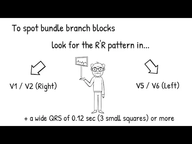

Easily remembered as: In English we read from left to right, but the ECG prints a view of the heart from right (V1-V2) to left (V5-V6).
Reference
Dubin, D. (2000) Rapid Interpretation of EKG’s. 6th edition. Florida: COVER Publishing Company.
Like it? Hate it? Spot a mistake? Let me know in the comments below ->
Check out the video summary below.
For more information, have a look at the related Article 999 post here.
Other posts that may interest you:
Fast Fact: Head Injury is the commonest cause of death
Reference: National Institute for Health and Care Excellence. (2014). Head injury: assessment and early management (NICE Clinical Guideline 176). Retrieved from https://www.nice.org.uk/guidance/cg176
This has not yet been peer reviewed. Comment and review below 🙂
This post shows the key points and quotes as they relate to frontline ambulance staff; anything not relevant has not been included, but you may read more by following the link to the guidelines here.
Remember to read our disclaimer before putting into practice anything you see, hear, or read here. Also remember to check your local guidelines and the scope of practice for your role before putting any of this into practice.
Article 999: Simplifying the Long Stuff; Presenting the Relevant Stuff; Refreshing you on the Important Stuff.
www.article999.co.uk

‘any trauma to the head other than superficial injuries to the face.’ p6
National Institute for Health and Care Excellence [NICE], 2014: 6
‘Head injury is the commonest cause of death and disability in people aged 1-40 years in the UK.’
NICE, 2014: 6
‘The incidence of death from head injury is low, with as few as 0.2% of all patients attending emergency departments with a head injury dying as a result of this injury.’
NICE, 2014: 6
‘the majority of fatal outcomes are in the moderate (GCS 9-12) or severe (GCS 8 or less) head injury groups’
NICE, 2014: 6

‘For adults who have sustained a head injury and have any of the following risk factors’
–> This should happen within one hour of identifying the situation.
(NICE, 2014: 10)
*Consider ‘the pre-injury baseline GCS may be less than 15. Establish this where possible’ (NICE, 2014: 19).
If the patient is on anticoagulants and has none of the above, they should have a CT head ‘within 8 hours of the injury’. (NICE, 2014: 12)

(NICE, 2014: 13)

Any of:
(NICE, 2014: 11)
If a child has ‘more than one’ of these, he/she should have a CT scan within an hour:
(NICE, 2014: 11)
Note: If the child has just one of these, he/she ‘should be observed for a minimum of 4 hours after the head injury’ and if he/she then develops more of the above, a CT is warranted. (NICE, 2014: 12)
The rules regarding CT cervical spine scans are different in children, compared to adults, due to the risk of radiation to their thyroid.


And:
Also, ‘depending on judgement of severity:’


During these transfers, patients ‘should be accompanied by a doctor with appropriate training and experience in the transfer of patients with acute brain injury. […] Patients requiring non-emergency transfer should be accompanied by appropriate clinical staff.’
NICE, 2014: 32-33

National Institute for Health and Care Excellence. (2014). Head injury: assessment and early management (NICE Clinical Guideline 176). Retrieved from https://www.nice.org.uk/guidance/cg176

#medicalwriting #nice #paramedic #emt #frontline #emergency #ambulance #headinjury
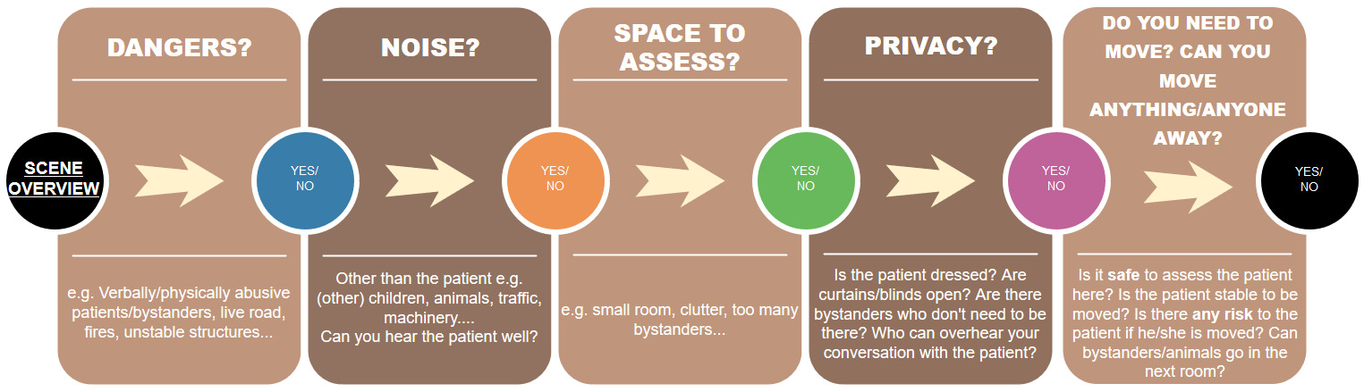
And more…

You may find varying suggestions for what to consider in what is usually referred to as your ‘scene assessment’ (Harris, 2016: 1; Pilbery & Lethbridge, 2016: 126) or ‘end of bed […] assessment’ (Spurr, 2014). This is a cheat sheet to assist you in identifying these important factors. There are a few examples within each category, forming part of what is surely an endless list. Harris (2016: 1-3) also recommends considering use of PPE, considering vehicle position, and considering CBRNE incidents. Pilbery & Lethbridge (2016: 127) recommend considering outside/inside temperature, terrain that is not reachable by land ambulance, and referring to major incident guides where applicable. Major incidents are not covered within this cheat sheet, but this sheet may guide you through the initial thought process prior to announcing a major incident…
Final Note & References
This is a cheat sheet which is designed to complement your scene assessment. This cheat sheet is in the author’s own words but ideas have been used from the below texts. Any copies of the above cheat sheets should be paired with the below references.
Harris, G. 2016. ‘General principles of assessment’ in A.Y. Blaber and G. Harris, ed. 2016. Assessment Skills for Paramedics, 2nd ed. Berkshire: Open University Press, pp. 1-13
Pilbery, R. and Lethbridge, K. 2016. Ambulance Care Practice, Bridgwater: Class Professional Publishing
Spurr, J. 2014. End-of-Bedogram: The Art Formally Known as Intuition, Available Online: http://injectableorange.com/2014/03/end-bedogram-art-formally-known-intuition/ (Accessed 26/03/18)
Feedback? Comments? Please post below…

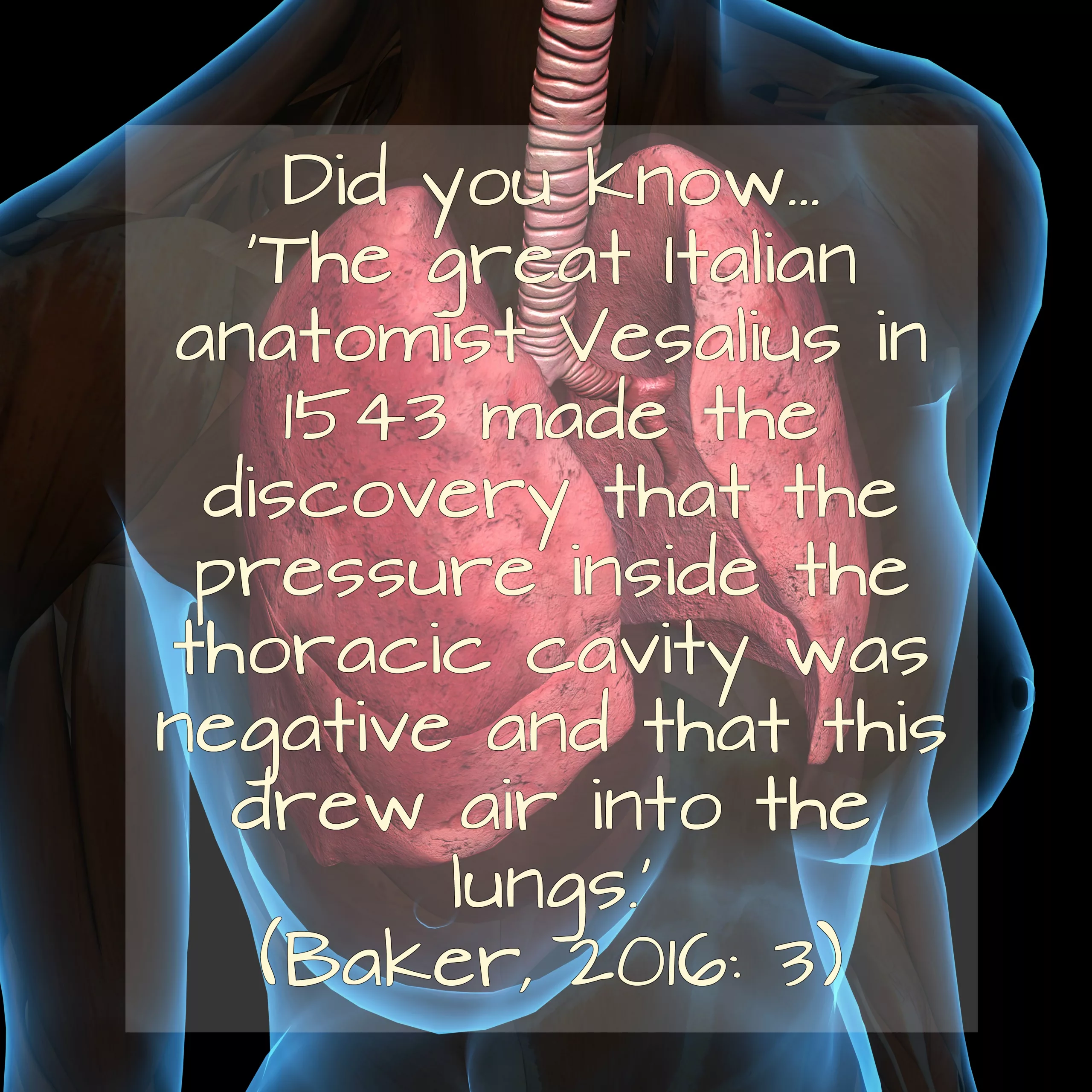
A shorter version of Ventilators: Why, What, How, When? featuring how-to use the ventilator demonstrations only. Made by UK Paramedics, this demonstrates how to use the PneuPac ParaPac ventilator.
Read more:
Ventilators: Why, What, How, When?
How do I get the ventilator off the racks?
What is the relief pressure dial on the ventilator?
The Discovery of Negative and Positive Pressure Ventilation – Fast Fact
What is positive pressure breathing?
What is Negative Pressure Breathing?
Disclaimer
You must read the full disclaimer at www.article999.co.uk/about/ (disclaimer tab) before putting into place anything you see here. Useful information is available in the text on this video, so if you only listen to the narration you will miss important facts. The ventilation settings mentioned in this video are what is recommended by Smiths Medical, current guidelines and some articles – however, as with all topics, there is always varying information available online & alternative expert advice, and no video can cater for all of that. Similarly, this video is intended to demonstrate how to use equipment & to introduce or remind you to the Why, What and When of ventilators – not to tell you that you should or shouldn’t be using it. That is dependent on local guidelines, your research & your choice as a clinician. This video merely highlights the varying advice regarding tidal volume settings, and in the text points out the potential problems with some of the figures. No specific volume is recommended.
Transcript
Article 999. Ventilators: Why, What, How, When?
This video has been made by UK Paramedics following guidelines. It is not endorsed by any author, organisation or Ambulance Trust. You must read the full disclaimer at www.article999.co.uk/about/ and refer to your local guidelines before putting into place anything you see here. This video is intended to demonstrate how to use the ventilator according to use guides and guidelines – not to tell you that you should or shouldn’t use it. That decision is up to you and should be dependent on local guidelines and your own research. This is what the textbooks, the manufacturer and a few articles say & is not intended to represent the expert opinions or experience of others within healthcare.
Remember hand hygiene, bare below the elbows and gloves in real life.
How?
(Gregory & Mursell, 2010: 44; Pilbery & Lethbridge, 2016: 189; Baker, 2012; Smiths Medical, 2017)
The following demonstrate the variance in researched texts and guidelines – not necessarily the advised settings:
| 400-600ml | Baskett, 1996 |
| 500-600ml | Perkins et al, 2015 |
| 600ml | Baker, 2016 |
| 800-900ml | Smiths Medical, 2017 – may not be advisable due to risks of barotrauma. See below |
| 6-8ml/kg | Bocklage & Balk, 2017; Frakes, 2007 |
| 10ml/kg or 5-8ml/kg | Baker, 2012 – depending on the extent of the risk of barotrauma |
7. Next, temporarily occlude the patient connection. The relief pressure monitor should go to the maximum setting – i.e. 40cmh20.
(Gregory & Mursell, 2010: 44; Pilbery & Lethbridge, 2016: 189; Baker, 2012; Smiths Medical, 2017)
What do the audible warnings mean?
(Smiths Medical, 2017)
High pressure = excessive tidal volume, incorrect airway position, kinked ET tube, or incorrect ventilation settings.
If the pressure is reaching it’s max, there may be something wrong with the circuit.
If you’ve checked the above and the alarm is still sounding, Smiths Medical (2017) advise that your tidal volume setting might simply be too high.
Low pressure = leakage or insufficient tidal volume/settings, faulty valve in the patient’s circuit
This alarm tends to occur when pressure drops below 10cmh20
Earlier, I pointed out the alarm in the middle, at the bottom. This is SMMV. It’s an indicator that will flash green if the patient is breathing for themselves. The ventilator will assist if the patient breathes with less than 150ml of tidal volume. Between 150-400ml the ventilator will extent the exhalation time to allow the patient to complete their own breath, and above 400ml the ventilator will allow the patient to breathe and will not assist, but may still assist on the next breath if required (Smiths Medical, 2017; Baker, 2012).
Extra facts:
McCarty et al (2012) found that ‘ventilation rates and tidal volumes commonly exceeded Guideline recommendations. This resulted in […] excessive mean airway pressure’
Smiths Medical (2017) advise that peak inflation pressure should be below 20cmh20 when using a mask to ventilate due to the issues of too high pressure
Inspiration to Expiration should be 1:2 (Smiths Medical, 2017; Baker, 2012)
References
Baker, D. 2012. Emergency and Transport Ventilation, an introductory guide, Smiths Medical International Limited: Luton
Baker, D.J. 2016. Artificial Ventilation: A Basic Clinical Guide. Springer International: Switzerland
Baskett, P. et al. 1996. ‘Tidal volumes which are perceived to be adequate for resuscitation’, Resuscitation, 31 (3), pp. 231-4
Bocklage, T. & Balk, R.A. 2017. Setting the Tidal Volume In Adults Receiving Mechanical Ventilation: Lessons
Learned From Recent Investigations, Available Online: https://www.nbrc.org/wp-content/uploads/2017/07/Setting-the-Tidal-Volume.pdf (Accessed 17/12/17)
Frakes, M. 2007. Ventilation Modes and Monitoring, Available Online: http://www.rtmagazine.com/2007/02/ventilation-modes-and-monitoring/ (Accessed 17/12/17)
Gregory, P. and Mursell, I. 2010. Manual of Clinical Paramedic Procedures, West Sussex: John Wiley & Sons
McCarty, K. et al, 2012. Ventilation rates and tidal volume during emergency department cardiac resuscitation, Resuscitation, 83: 4, p45
Owen, R. and Castle, M. 2006. ‘EtCO2: the key to effective prehospital ventilation’, Emergency Medical Journal, 23 (7), pp. 578-579
Perkins, G.D. 2015. European Resuscitation Council Guidelines for Resuscitation 2015: Section 2. Adult basic life support and automated external defibrillation, Available Online: https://ercguidelines.elsevierresource.com/european-resuscitation-council-guidelines-resuscitation-2015-section-2-adult-basic-life-support-and/fulltext#back-bib0760 (Accessed 17/12/17)
Pilbery, R. & Lethbridge, K. 2016. Ambulance Care Practice, Bridgwater: Class Professional Publishing
Smiths Medical, 2017. ‘Pneupac – ParaPAC’, Pneupac Transport Ventilators – Breathe Easy, Available Online: https://www.smiths-medical.com/resources/pneupac-transport–ventilators—breathe-easy (Accessed 27/10/17)
With thanks to Smiths Medical for their correspondence and information
Music:
Dobroide, 2010. 20091229.ambulance.siren.wav. Available Online: https://freesound.org/people/dobroide/sounds/8713/ (Accessed 21/08/17)
Productiontrax.com
Images:
Article 999’s own or licenses purchased via Adobe Stock & Dreamstime

If you’ve ever felt the frustration of being a student on Day 1 and having to retrieve the ventilator for your crewmate, you might enjoy this GIPHY.