Category: summary

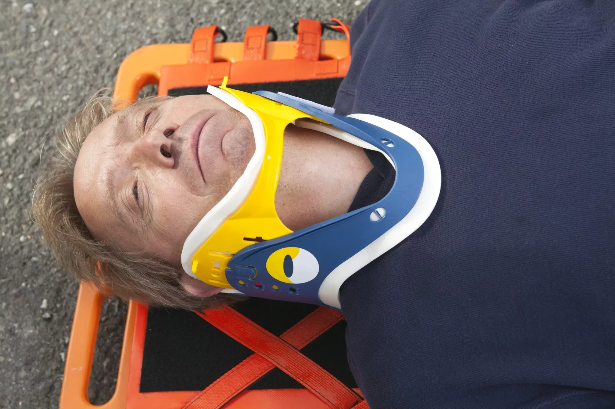
  • What does NICE say about head injuries?

    What does NICE say about head injuries?

    This post is based on the NICE Clinical Guidelines, 2014. One part of their guidelines was in turn updated in 2019.

    This post shows the key points and quotes as they relate to frontline ambulance staff; anything not relevant has not been included, but you may read more by following the link to the guidelines here.

    Remember to read our disclaimer before putting into practice anything you see, hear, or read here. Also remember to check your local guidelines and the scope of practice for your role before putting any of this into practice.

    Article 999: Simplifying the Long Stuff; Presenting the Relevant Stuff; Refreshing you on the Important Stuff.

    www.article999.co.uk

    Definition of Head Injuries

    ‘any trauma to the head other than superficial injuries to the face.’ p6

    National Institute for Health and Care Excellence [NICE], 2014: 6

    Statistics

    ‘Head injury is the commonest cause of death and disability in people aged 1-40 years in the UK.’

    NICE, 2014: 6

    ‘The incidence of death from head injury is low, with as few as 0.2% of all patients attending emergency departments with a head injury dying as a result of this injury.’

    NICE, 2014: 6

    ‘the majority of fatal outcomes are in the moderate (GCS 9-12) or severe (GCS 8 or less) head injury groups’

    NICE, 2014: 6

    When might adults need to attend A&E for a CT head scan following a head injury?

    ‘For adults who have sustained a head injury and have any of the following risk factors’

    • Reduced GCS, ‘less than 13’ initially, or ‘less than 15 at 2 hours after the injury on assessment in the emergency department’*
    • suspected skull fracture of any type
    • ‘post-traumatic seizure’
    • Neurological deficit
    • ‘More than 1 episode of vomiting’

    –> This should happen within one hour of identifying the situation.

    (NICE, 2014: 10)

    *Consider ‘the pre-injury baseline GCS may be less than 15. Establish this where possible’ (NICE, 2014: 19).

    If the patient is on anticoagulants and has none of the above, they should have a CT head ‘within 8 hours of the injury’. (NICE, 2014: 12)

    When might adults need to attend A&E for a CT cervical spine following a head injury?

    • ‘The patient has been intubated’
    • ‘The patient is having other body areas scanned for head injury
    • ‘…there is clinical suspicion of cervical spine injury and any of the following apply:
      • => 65
      • ‘dangerous mechanism of injury’
      • neuro deficit
      • ‘paraesthesia in the upper or lower limbs’

    (NICE, 2014: 13)

    When might children need to attend hospital for a CT head scan following a head injury?

    Any of:

    • ‘Suspicion of non-accidental injury’
    • ‘Post-traumatic seizure but no history of epilepsy’
    • Reduced GCS <14 initially, <15 2 hours later
    • For under 1 year olds, Reduced GCS <15 on the paediatric scale
    • Suspected skull fracture of any type
    • Neuro deficit
    • For under 1 year olds, ‘presence of bruise, swelling or laceration of more than 5 cm on the head’

    (NICE, 2014: 11)

    If a child has ‘more than one’ of these, he/she should have a CT scan within an hour:

    • ‘Loss of consciousness lasting more than 5 minutes (witnessed)’
    • ‘Abnormal drowsiness’
    • Vomiting x3 or more episodes
    • ‘Dangerous mechanism of injury (high-speed road traffic accident either as pedestrian, cyclist or vehicle occupant, fall from a height of greater than 3 metres, high-speed injury from a projectile or other object)’
    • ‘Amnesia […] lasting more than 5 minutes’

    (NICE, 2014: 11)

    Note: If the child has just one of these, he/she ‘should be observed for a minimum of 4 hours after the head injury’ and if he/she then develops more of the above, a CT is warranted. (NICE, 2014: 12)

    The rules regarding CT cervical spine scans are different in children, compared to adults, due to the risk of radiation to their thyroid.

    ‘Consider or suspect abuse as a contributory factor to or cause of head injury in children’ (NICE, 2014: 7)

    For what other reasons should an adult or child with a head injury attend A&E?

    • loss of consciousness
    • ‘Amnesia for events before or after the injury’
    • ‘Persistent headache since the injury’
    • ‘Any vomiting episodes since the injury’ – but NICE advise considering the causes of single vomiting episodes in those under 12
    • ‘Any seizure since the injury’
    • ‘Any previous brain surgery’
    • ‘A high-energy head injury’
    • ‘Any history of bleeding and clotting disorders’
    • Anticoagulants
    • ‘Current drug or alcohol intoxication’
    • Safeguarding issues
    • ‘Continuing concern by the professional about the diagnosis’
      (NICE, 2014: 17-18)

    And:

    • Patients who, 48 hours later, have ‘any persistent complaint relating to the initial head injury’
      (NICE, 2014: 23)

    Also, ‘depending on judgement of severity:’

    • ‘irritability or altered behaviour’
    • Other ‘Visible trauma to the head […] of concern to the professional’
    • ‘No one is able to observe the injured person at home’
    • ‘Continuing concern by the injured person or their family or carer about the diagnosis’
      (NICE, 2014: 18)

    What else should I consider in my assessment and treatment of a patient with a head injury?

    • For adults, NICE recommends ‘managing their care according to clear principles and standard practice’ as in the ATLS and PHTLS courses, and the JRCALC for adults, and the APLS and PHPLS courses for children. There are others referenced, but Article 999 has included the most relevant here.
      (NICE, 2014: 19-20)
    • ‘Manage pain effectively because it can lead to a rise in intracranial pressure’
      (NICE, 2014: 21)
    • ‘Ascribe depressed conscious level to intoxication only after a significant brain injury has been excluded’
      (NICE, 2014: 21-22)
    • Pre-alert patients with a reduced GCS, especially of <8. They will most likely need anesthetist or critical care involvement
      (NICE, 2014: 21-22)

    What’s worth bearing in mind during hospital-neuroscience unit transfers of patients with head injuries?

    • Patients who have a GCS of less than 8 should be intubated
    • Before transporting, make sure to stabilise the patient and ensure monitoring is attached
    • A patient ‘with persistent hypotension’ should not be transported until they are ‘stabilised’
      (NICE, 2014: 32-33)

    During these transfers, patients ‘should be accompanied by a doctor with appropriate training and experience in the transfer of patients with acute brain injury. […] Patients requiring non-emergency transfer should be accompanied by appropriate clinical staff.’

    NICE, 2014: 32-33

    Reference

    National Institute for Health and Care Excellence. (2014). Head injury: assessment and early management (NICE Clinical Guideline 176). Retrieved from https://www.nice.org.uk/guidance/cg176

    Article 999: Simplifying, Presenting, Refreshing

    #medicalwriting #nice #paramedic #emt #frontline #emergency #ambulance #headinjury

  • PARAMEDIC-2 Trial Results

    PARAMEDIC-2 Trial Results


    ‘For more than 50 years, treatment strategies have included the use of various drugs, but there is limited evidence that such treatments are effective’ (Perkins et al, 2018).

    Now the PARAMEDIC-2 trial results are in…

    • 8014 patients of the 10, 623 initially selected were included in the trial.
    • Of these, 4015 were given adrenaline and 3999 a placebo.
    • Of those who were given adrenaline and survived until discharge, 39 (31.0%) out of 126 patients had a ‘severe neurological impairment’ and of those who were given the placebo, the same was true for ’16 of 90 patients [17.8%]’
      (Perkins et al, 2018).

    Exclusions to the trial

    • ‘…those with anaphylaxis, pregnant women and children’ (below age 16).
    • After the pilot, life-threatening asthma as a suspected cause of the cardiac arrest was added as an exclusion because of ‘the potential overlap between the presentation of asthma and anaphylaxis’.
    • Anaphylaxis was excluded because adrenaline is considered potentially ‘beneficial’ for this condition
      (Warwick Clinical Trials Unit, 2018).

    The trial only looked at the effects of the use of adrenaline during out-of-hospital cardiac arrest and not at the use of adrenaline during ROSC, which is a protocol that some Trusts follow (Warwick Clinical Trials Unit, 2018). In addition, this does not cover any treatment initiated by the hospital if the patient was transported there.

    Conclusions

    ‘the benefits of epinephrine that were identified in our trial are small, since they would result in 1 extra survivor for every 112 patients treated. This number is less than the minimal clinically important difference that has been defined in previous studies.29,30 Among the survivors, almost twice the number in the epinephrine group as in the placebo group had severe neurologic impairment’ (Perkins et al, 2018).

    Limitations

    Perceived limitations include: ‘Information about the quality of CPR was limited to the first 5 minutes of cardiac arrest and involved fewer than 5% of the enrolled patients.’ (Perkins et al, 2018)

    Further limitations to consider could be:

    • time to CPR
    • patient’s co-morbitities that weren’t already considered in the study

    More information

    Warwick’s Clinical Trials Unit have produced an infographic with more information here.

    View the University of Warwick’s press release here.

    References

    NIHR, 2018. Investigating the role of adrenaline in cardiac arrest. Available Online: https://www.nihr.ac.uk/news/investigating-the-role-of-adrenaline-in-cardiac-arrest/8931 (Accessed 19/07/18)

    Perkins, G.D. et al, 2018. A randomized trial of epinephrine in out-of-hospital cardiac arrest. New England Journal of Medicine. doi: 10.1056/NEJMoa1806842

    Warwick Clinical Trials Unit, 2018. Available Online: https://warwick.ac.uk/fac/med/research/ctu/trials/critical/paramedic2/faqs/ (Accessed 19/07/18)


  • Cheat Sheets: Scene Overview

    Scene Overview

    Harris, 2016; Pilbery & Lethbridge, 2016 *This can be made available in alternative formats & colours on request*

    And more…

    Harris, 2016; Pilbery & Lethbridge, 2016 *This can be made available in alternative formats & colours on request

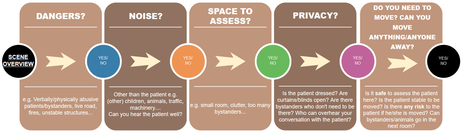
    You may find varying suggestions for what to consider in what is usually referred to as your ‘scene assessment’ (Harris, 2016: 1; Pilbery & Lethbridge, 2016: 126) or ‘end of bed […] assessment’ (Spurr, 2014). This is a cheat sheet to assist you in identifying these important factors. There are a few examples within each category, forming part of what is surely an endless list. Harris (2016: 1-3) also recommends considering use of PPE, considering vehicle position, and considering CBRNE incidents. Pilbery & Lethbridge (2016: 127) recommend considering outside/inside temperature, terrain that is not reachable by land ambulance, and referring to major incident guides where applicable. Major incidents are not covered within this cheat sheet, but this sheet may guide you through the initial thought process prior to announcing a major incident…

    Final Note & References

    This is a cheat sheet which is designed to complement your scene assessment. This cheat sheet is in the author’s own words but ideas have been used from the below texts. Any copies of the above cheat sheets should be paired with the below references.

    Harris, G. 2016. ‘General principles of assessment’ in A.Y. Blaber and G. Harris, ed. 2016. Assessment Skills for Paramedics, 2nd ed. Berkshire: Open University Press, pp. 1-13

    Pilbery, R. and Lethbridge, K. 2016. Ambulance Care Practice, Bridgwater: Class Professional Publishing

    Spurr, J. 2014. End-of-Bedogram: The Art Formally Known as Intuition, Available Online: http://injectableorange.com/2014/03/end-bedogram-art-formally-known-intuition/ (Accessed 26/03/18)


    Feedback? Comments? Please post below…

    This post is yet to be peer reviewed. Please get in touch if you have any comments.

  • How do I get the ventilator off the racks?

    How do I get the ventilator off the racks?

    If you’ve ever felt the frustration of being a student on Day 1 and having to retrieve the ventilator for your crewmate, you might enjoy this GIPHY.


  • What is the relief pressure dial on the ventilator?

    What is the relief pressure dial on the ventilator?

     This article refers to the workings of Smith’s Medical Pneupac ParaPac and the newer ParaPac Plus. Other transport ventilators may vary in their settings and processes. Please check what unit your ambulance trust has before following the information here.


    [tabby title=”Detail”]

    The pressure of air inside our lungs is normally just right to not only ensure that negative pressure breathing occurs but also to prevent a shunt (where blood bypasses the lungs) (Hess & Kackmarek, 2014: 431), the oesophageal spinchter from opening, causing air to enter the stomach (Baker, 2012: 1), and a host of other potential problems. Any means of supported ventilation where the patient is not breathing for themselves produces positive pressure ventilation. Not only does this change the way air enters the patient’s lungs while they are ventilated, but it also comes with all of the above risks if any of the settings are incorrect, and more.

    Many of the effects ‘are related to mean airway pressure [which is] […] the average pressure applied to the airway during the ventilatory cycle’ (Hess & Kackmarek, 2014: 420) With too ⇑ pressure comes ⇓ venous return (Hess & Kackmarek, 2014: 420) and ⇑ chance of barotrauma (Baker, 2016: 75).

    To summarise, the potential negative effects are:

    • Blood bypassing the lungs (a shunt) (Hess & Kackmarek, 2014: 431)
    • Air entering the stomach (Baker, 2012: 1)
    • Venous return decreasing (Hess & Kackmarek, 2014: 420)
    • Barotrauma (Baker, 2016: 75)

    Because modern ventilators are volume-controlled, preventing a different problem that occurs during pressure-controlled ventilation, there is a set tidal volume which the ventilator will always provide to the patient. To provide this volume, the pressure will constantly increase until the volume is reached (Baker, 2016: 132).

    The Means of Avoiding These Problems

    In ventilators, the pressure of an oxygen cylinder ‘is lowered through a reducing valve down to 30-90psi’ from 2000psi. It’s then delivered to the patient ‘to a maximum of 40cmh20 or 60cmh20’, (Baker, 2012: 18)*, depending on whether you’re in the UK or the US.

    *cmh20 = centimetres of water pressure (SensorsONE, 2017)

    Since we can’t have too much pressure there needs to be a way of stopping the ventilator if things get awry. The relief pressure is what this refers to. Via a safety valve (Baker, 2012), otherwise known as ‘exhaust valves’ the supply is cut and excess peak inspiratory pressure (PIP) is released into the atmosphere (Zaconeta et al, 2010), ‘prevent[ing] over-ventilation of the lungs’ (Baker, 2016: 145)

    Why would the pressure get this high anyway?

    • ‘The patient circuit […] [could be] blocked or kinked’ (Baker, 2016: 142). Best to check it.
    • ‘If the lung compliance[*] is low and the airway resistance is high the pressure can rise to high levels’ because, as above, the pressure will keep rising until the set tidal volume is delivered (Baker, 2016: 142).
    • The wrong relief pressure settings.

    *Compliance ‘refers to the ease of expansion of the lungs and the walls of the thorax’ (Baker, 2012: 8). Ordinarily, according to Baker (2012: 8), the value is 100 cmh20 but when ventilated it’s 40-50cmh20. Physical injuries and medical conditions may further alter this figure.

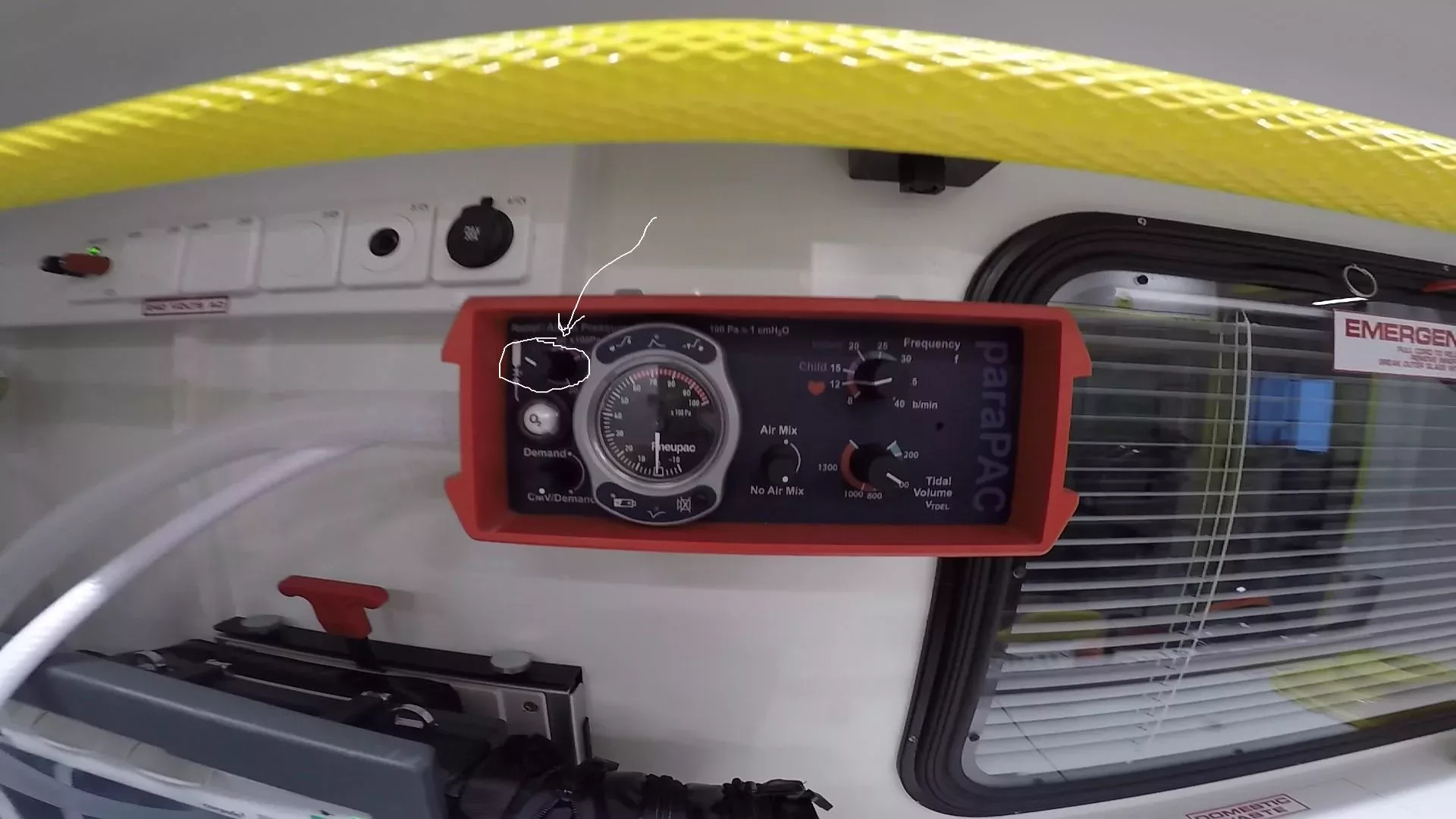
    Which dial are you talking about?

    This one

    The Pneupac ParaPac (Smiths Medical). Photo highlights the Relief Pressure dial.

    What should I set it to?

    40cmh20 if you’re in the UK (Baker, 2012)

    Why is the relief pressure set at 40cmh20 in the UK? Why is it 60cmh20 in the US?

    Article 999 is struggling to find the answers to these questions. If you know the answers, please get it touch. It would be great to add the information to this resource.

    This post is yet to be peer reviewed. Please get in touch if you have any comments.


    [tabby title=”Super Summary”]

    Article 999 previously posted an article on positive pressure ventilation, including its risks. The aim of positive pressure ventilation is to ‘maximise the potential benefit of mechanical ventilation while minimizing the potential for harm’ (Hess & Kackmarek, 2014: 407). The relief pressure dial on the ventilator ensures that the pressure of ventilation is not so high as to cause blood bypassing the lungs (Hess & Kackmarek, 2014: 407), the oesophageal spinchter opening (Baker, 2012: 1), barotrauma (Baker, 2016: 75; Hess & Kackmarek, 2014) and more. The ventilator’s other settings ensure that the patient actually receives ventilation, but the relief pressure setting ensures that a valve drains excess air when settings, poor setup, or the patient’s lung compliance (Baker, 2016: 43) have caused the pressure to get to high.

    What should I set it to?

    40cmh20 (Baker, 2012)

    What should I look out for if the pressure is too high?

    Kinked tubing (Baker, 2016: 142) & your settings

    Read more in the Detail tab.

    [tabby title=”Prefer to see a diagram? Check out the mind-map summary”]


    [tabby title=”References”]

    References

    Baker, D. 2012. Emergency and Transport Ventilation, an introductory guide, Smiths Medical International Limited: Luton

    Baker, D.J. 2016. Artificial Ventilation: A Basic Clinical Guide. Springer International: Switzerland

    Hess, D.R. & Kacmarek, R.M. 2014. Essentials of Mechanical Ventilation. McGraw Hill Education: New York

    SensorsONE, 2017. cmh20 – Centimetres of Water Column at 4 deg C Pressure Unit, Available Online: https://www.sensorsone.com/cmh2o-centimetres-water-column-4-deg-c-pressure-unit/ (Accessed 16/11/17)

    Zaconeta, C.A.M. et al, 2010. ‘Evaluation of peak inspiratory pressure and respiratory rate during ventilation of a preterm infant lung model with a self-inflating bag by paramedics of the Fire Department’, Revista Paulista de Pediatria, vol. 28. No. 1.

    Not endorsed by but with thanks to Smiths Medical for providing information.

    [tabbyending]


  • Should We Use Rigid Cervical Collars?

    Should We Use Rigid Cervical Collars?

    [table id=2 responsive=scroll datatables_buttons=”colvis,copy,csv,excel,pdf,print” /]


    Peer reviewed?


    This post is due to be peer reviewed. All of Article 999’s posts will soon be updated with this image, or with an image that shows 1 or more, or 10 or more individuals have peer reviewed that post.


  • What are the main sections used for?

    What are the main sections used for?

    [tabby title=”This tab: 3) What are the main sections used for?” open=”yes”]

    Section 2 Purpose: Admission for patient assessment.

    Duration of section: 28 days


    Section 3 Purpose: Admission for treatment of a patient.

    Duration of section: up to 6 months


    Section 4 Purpose: Urgent admission for patient assessment from the community, usually enforced when a section 2 would take to long to enact.

    Duration of section: 72 hours (may be converted to section 2 to extend assessment period to 28 days)


    Section 5(2) Purpose: Urgent detention of inpatient

    Duration of section: 72 hours


    Section 5(4) Purpose: Urgent detention of an inpatient by a nurse were a doctor is absent

    Duration of section: 6 hours


    Section 135 Purpose: Removal of person from home to place of safety. This requires a court order to enact and remove someone from a private property.

    Duration of section: 72 hours


    Section 136 Purpose: Removal of a person from a public place to a place of safety. This does not require a court order.

    Duration of section: 72 hours


    Community Treatment Order Purpose: An order that a patient is placed upon after discharge from hospital for psychiatric treatment that allows continued treatment within the community setting.

    Duration of section: up to 6 months.

    References

    • Crown Copyright (1983) Mental Health Act 1983 [online]. Available at: http://www.legislation.gov.uk/ukpga/1983/20/contents (Accessed 21/07/17) (amended by the Mental Health Act 2007)
    • J. Collier, M. Longmore, T. Turmezel and A. R. Mafi. Oxford Handbook of Clinical Specialties, 8th edition, Oxford University press, 2008, pp: 398-401

    [tabbyurl title =”1. What is a section?” url=”https://article999.co.uk/what-is-a-section” indicator=”ext”]

    [tabbyurl title=”2. Who can enforce a sectioning order?” url=”https://article999.co.uk/can-enforce-sectioning-order” indicator=”ext”]

    [tabbyending]

  • What is a section?

    What is a section?

    [tabby title=”1) What is a section?” open=”yes”]

    A sectioning order is legislation powers given to health care professionals and police officers under the Mental Health Act’s of 1983 and 2007 that allow for compulsory admission of an individual to hospital or a place of safety. These are enacted if a patient is judged to have a mental disorder that is sufficiently severe to require treatment for said disorder or to remove a person to a place of safety who could be at risk of being a danger to themselves or the public.


    References:

    • Crown Copyright (1983) Mental Health Act 1983 [online]. Available at: http://www.legislation.gov.uk/ukpga/1983/20/contents (Accessed 21/07/17) (amended by the Mental Health Act 2007)
    • J. Collier, M. Longmore, T. Turmezel and A. R. Mafi. Oxford Handbook of Clinical Specialties, 8th edition, Oxford University press, 2008, pp: 398-401

    [tabbyurl title=”2. Who Can Enforce a Sectioning Order?” url=”https://article999.co.uk/can-enforce-sectioning-order” indicator=”ext”]

    [tabbyurl title= “3. What are the main sections used for?” url=”https://article999.co.uk/main-sections-used” indicator=”ext”]

    [tabbyending]


  • Who can enforce a sectioning order?

    Who can enforce a sectioning order?

    [tabby title=”2. This tab: Who can enforce a sectioning order?” open=”yes”]

    There are several different types of sectioning orders as listed in the relevant tabs. Each one is used for different purposes, lasts for different lengths of time and can be used by a variety of different health care professionals and police officers. However, not everyone may use every type of order. The following are the sectioning powers available to the following professions:


    Nurses: Section 5 (4)
    Nurses: Section 5 (4)


    Doctors: Section 5 (2)
    Doctors: Section 5 (2)


    Police Officers: Sections 135 and 136
    Police Officers: Sections 135 and 136


    A mental health professional and doctor:
    A mental health professional and doctor:

    Approved mental health professionals (social workers, nurses, psychologists or occupational therapists) and rarely relatives may also put an application in for sections 2, 3 and 4. These all require approval by 1 or more doctors to be carried out.


    References

    • Crown Copyright (1983) Mental Health Act 1983 [online]. Available at: http://www.legislation.gov.uk/ukpga/1983/20/contents (Accessed 21/07/17) (amended by the Mental Health Act 2007)
    • J. Collier, M. Longmore, T. Turmezel and A. R. Mafi. Oxford Handbook of Clinical Specialties, 8th edition, Oxford University press, 2008, pp: 398-401

    [tabbyurl title=”1. What is a section?” url=”https://article999.co.uk/what-is-a-section” indicator=”ext”]

    [tabbyurl title=”3. What are the main sections used for?” url=”https://article999.co.uk/main-sections-used” indicator=”ext”]

    [tabbyending]

  • Article Summary: Maxillofacial Trauma Patient

    Article Summary: Maxillofacial Trauma Patient

    An summary of a Krausz et al (2009) article discussing the importance of effective airway management in the maxillofacial trauma patient and the complexities that such an injury presents. Only points relevant to UK paramedics have been included. For more details, please read the original article. Any additions made by Article 999 are in square brackets [].

    [restabs alignment=”osc-tabs-left” pills=”nav-pills” responsive=”false” tabcolor=”#6ad61d”]
    [restab title=”Full Summary and Reference” active=”active”]

    The Key Points

    • Remember to follow ATLS protocol [and/or your local guidance and JRCALC guidelines]
    • Use cervical spine control along with effective airway management
    • ‘The most common critical care errors are related to airway and respiratory management. Gruen et al studied 25 trauma mortality patients […] [and] found that failure to intubate, secure or protect the airway was […] responsible for 16% of inpatient deaths’ [and that was in a trauma centre, albeit in 1996-2004!] (Gruen et al, 2006)

    Hutchinson et al (1990) (in Krausz et al, 2009) found 6 potential ways maxillofacial trauma might prevent effective airway management.

    These, from the top of the head downwards, can be summarised as:

    Head and soft tissue trauma
    – These risk ‘delayed airway compromise’

    Nasal bleeding/open wounds causing obstruction

    Mandible fractures
    – Leading to the tongue to drop back, blocking the oropharnyx
    – Leading to blockage of the nasopharnygeal airway

    Mouth obstructions
    – From loose items & bodily fluids

    Tracheal trauma
    – Leading to swelling and displacement of essential airway structures behind them.

    C-spine injury
    – Leading to the need for “in-line stablization”. This can cause a reduced view during intubation
    – The act of intubating might increase neck movements, potentially worsening this injury

    Stomach
    – [All patients ambulance personnel intubate in the prehospital environment might realistically have a full stomach]
    – Note the risk of regurgitation
    – Consider cricoid pressure – but also consider that this might ‘worsen the larnygeal view’
    – Other tips noted in this section are not relevant to UK paramedics. Please read the full article for more information

    These cause:
    – Difficulties fitting a mask
    – Less ‘efficient air transferring from the mask to the lungs’
    – A ‘difficulty in visualizing the vocal cords’ when intubating due to fluids & obstructions

    Final points

    • Consider all of the above and ‘avoid future complications’. Then address other injuries.
    • Emergency intubation is fraught with risks
    • Consider your expertise and experience. Ensure the trauma patient can access ‘the most experienced personnel’ where possible. This would reduce one of the risks.
    • Ensure prompt treatment
    • Consider the patient’s GCS, breathing level & risk to aid your decision making about transport
    • Check the ‘extent, the composition and the anatomy of the injury’. Is it possible to ventilate with a mask?
    • ‘Is there a limitation in mouth opening? Is that limitation the result of pain’ only? [In a prehospital environment, this may be difficult to ascertain]
    • An LMA may ‘not be suitable for managing trauma patients. However, it could enable ventilating the patient until definitive airway is achieved’

    References

    Gruen, R.L. et al. 2006. Patterns of Errors Contributing to Trauma Mortality: Lessons Learned from 2594 Deaths, Annals of Surgery, 244 (3): 371-380, Available Online:
    https://www.ncbi.nlm.nih.gov/pmc/articles/PMC1856538/

    Krausz, A. et al. 2009. Maxillofacial trauma patient: coping with the difficult airway, World Journal of Emergency Surgery, 4: 21, Available Online: https://wjes.biomedcentral.com/articles/10.1186/1749-7922-4-21

    [/restab]
    [restab title=”Article 999 Super Summary”]

    Remember to follow ATLS protocol [and your local guidelines], use cervical spine control and focus on airway management.

    Head and soft tissue trauma, nasal bleeding or open wounds, mandible fractures, mouth obstructions, tracheal trauma, c-spine injury and the patient’s stomach contents can all make airway management more challenging (Hutchinson et al, 1990 in Krausz et al, 2009).

    Top tips?

    • Consider cricoid pressure but bear in mind it may actually worsen your view during intubation.
    • Ensure rapid transport and treatment of the patient and consider your own expertise.
    • What level of experience do you really need to deal with this patient, who has it, and where are they?
    • The patient’s GCS, breathing level and risk should all support this decision.
    • You can use an LMA, but it’s a time-stop measure [Article 999 interpretation; see full summary].
    • Consider the ‘extent’ of the patient’s injuries and how they are going to make it difficult to use a mask and intubate if required.

    (Krausz et al, 2009)

    [/restab]
    [/restab][/restabs]