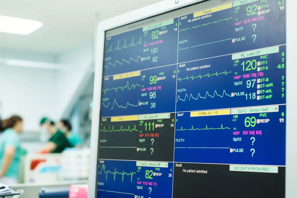
Sometimes I forget, so easily, what I’ve seen and who I’ve met, as if all of those experiences have accumulated into one. There is a beauty to this work – a sort of study of the potential of humankind, a sociological survey in a way, a means to observe the route to each eventuality.
We each explore every inch of our own naivety in the living conditions of every human and their pet, and the desperate circumstances that each person – regardless of their income – finds themselves in when they call 999: from the first cut finger that sends them into a panic, to the teenager whose first meal in days is too late to prevent a syncope. At the same time, we question, moan, degrade the lack of “common sense” in our neighbours. Or perhaps we degrade ourselves – after all, what has our common sense become after excessive exposure to illness and injury? What is our impression of “emergency” after years of practicing urgency in precise circumstances?
Perhaps, in our roles and years of experience, we have experienced growth upon growth – our pre-existing sensibility has been both nurtured and strained. We never did shout “emergency” for our own minor injuries before. Maybe that is what led us to work in the field we work within: we know to be calm when our legs can still take us to help, or we have adventured into such faraway places that we have had to find calm to find help – or worse, wait for pain to subside and injuries to heal without a polypharmacy. After all, this work attracts certain people who grit teeth rather than scream.
This work does more than that. It also reminds us that no matter how many days our washing is piling, no matter how long we have waited to clean the dishes, no matter how grubby our carpet, and no matter how strong the stench in the communal hallways, it could always be worse. There are homes in which I have tried tip-toeing (and failed) (you know, the ones that, following your visit, you see reason to stand on the Clinell Wipes you have just neatly placed on the ambulance floor; the ones that make you think, oh, that’s what those little plastic shoe coverings are really for), places in which I have had to wipe my glasses when I step inside, ones in which I have asked if I could open windows (and ones in which windows do not open). There are places in which we have hurried the grab bag back outside to prevent placing it on the infested floor, and ones we’ve immediately asked the patient to step away from.
Of course, this work has caused more. There are also places of inspiration – places in which I make a mental note of wall colours and furniture, places in which we admire the garden, and ones patients are so proud of they won’t leave, even during fires.
It’s not all material. There are patients who have proved to me the importance of healthy lifestyles and adaptable homes, ones who are so debilitated by conditions that they creak like an old tree – yet it is the patients much older who repeat endlessly, “never grow old.” Some have a point. Others are so disproven by age-defying marathon runners and glass half-filled storytellers that my empathy is reduced to a mere ‘hmm’ and a raise of my eyebrows.
The purpose of this blabbering is to remind myself, perhaps all of us, that the route to hospital can be an insightful one, as can the route home on a flight. Sometimes we forget what we see and who we meet. We forget how our paths intertwine, how a few different life circumstances or decisions could have led us to opposing roads. We work (or worked) so intensely, especially overnight, and the “I pay my taxes” or cut finger is too starkly followed by a cardiac arrest (or vice versa). Therefore, to work in this field is to see the juxtaposition of life and have it become normality, to the point we forget how much we have seen, as if it has all become one bubble, which perhaps occasionally we ought to let trickle.
This is your invitation to realise what you have seen and the characters you have met (ensuring confidentiality) – let the bubble of stories, of life, of potential possibilities trickle out, rather than float into the abyss.
“At Pentland Medical, we are proud to introduce innovative new products to the UK healthcare system. Hemashock not only has the potential to save lives but improve the longer term outlook for survivors of cardiac arrest and severe shock. We are excited to be part of this journey and show that there is a better way which will lead to better outcomes for patients” – Jamie Munro, Pentland Medical
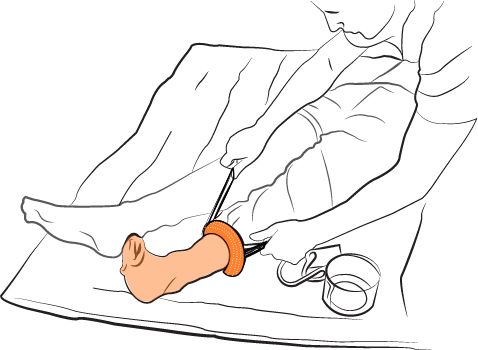
A new product in the UK market, the Hemashock from Pentland Medical, acts as an ‘autotransfusion tourniquet’ in cardiac arrest and hypovolemic shock. Rather than merely working as a tourniquet, these auto-transfusion tourniquets ‘sends blood from the legs to the core’. Here’s more about how this specific product works:
And here’s how it could look in practice for a solo-rescuer application (graphic only):
There is some limited evidence of the positive impact of this device. In one case of a PPH:
‘IV fluids and Pitocin 20 mg IV had no effect on bleeding or BP. An auto-transfusion tourniquet (A-TT), (HemaShock, OHK Medical Devices, Tirat Carmel, Israel) was placed on one of her legs. Within 1 minute blood pressure started to increase and heartrate slowed.’
Another case links survival from a presumed cardiac etiology to the application of this device. This is of course difficult to prove, and there are a number of variables that could have impacted the positive outcome – not just to ROSC (CPR quality, identification and/or treatment of other reversible causes, number of shocks) but also to discharge – in particular, target temperature management was used, something still under study and recently removed from Resuscitation Council guidelines following a lack of consistent evidence for its use. However, the article certainly reads convincingly – a patient who is in refractory VF, who swiftly develops ROSC following application of the device.
In fact, in a clinical validation at one medical centre, the device appears to have resulted in a higher chance of ROSC in cardiac arrest patients who have had prolonged resuscitation and poor indicators for survival. Notably though, only one patient survived to discharge.
For further medical evidence relating to the product, please follow this link to the product page.
Of course, application of any new product comes with barriers, and as with most interventions we must ask if it is not only necessary but also if it causes any delay or depreciation in quality of the basic and most effective interventions in a medical arrest. This study investigated the use of auto-transfusion tourniquets during cardiac arrest and found that an effective team can deploy them without impacting CPR quality.
Limitations:
There are currently no large-scale clinical trials of this device, and the device does not appear to have been tested independently of other variables e.g. target temperature management in ROSC, which is another experimental area given its addition and subsequent removal from the Resuscitation Council guidelines. Additionally, despite auto transfusion tourniquets being investigated in the study above from a team-working perspective, the actual speed of application does not appear to have been examined, something which has been heavily investigated in ordinary tourniquets. Additional human factor elements should be considered here, too – the minimum staff numbers required for application, and the minimum training required for efficient application are both high-stake areas that require further investigation.
However, the product remains an intriguing dual-purpose device with demonstrable results, and one that adds a positive vision to the future of cardiac arrest management and hypovolemia.
Summary:
Please note, this article has been produced by a request from Innovel Medical. No financial or other incentive beyond mere site promotion has been involved, no practical exposure to the product mentioned has been experienced, and this article does not constitute a product recommendation. As always, Article 999 is disconnected from any employers the author works for (see the full disclaimer via the site menu).
MONA (Morphine, Oxygen, Nitrates, Aspirin), has been a long-taught concept for ACS management (Kline, Conti, and Winchester, 2015). Though listed in reverse order and not summarised by an acronym, the same treatment list remains in Resuscitation Council ILS course slide content, and each one has ‘if required’ written in brackets – a key point when two out of four treatments have contra-indications and one is a feature of at least variable evidence. Throughout my development as an ALS instructor, I have learned to encourage giving oxygen early in a patient assessment, before reliable saturations can be gained, or ideally Pac02 readings because this can prevent early deterioration of unwell patients. There are often candidates who have heard of MONA before, or recall treatments in that order – yet I have also come across people who adamantly refuse to teach it because they disagree with oxygen being so heavily emphasised. Is MONA no longer advised – despite each element of MONA still being given? Are we to tell those familiar with the acronym that it is old news and outdated practice? I have looked into the research in order to understand this better. Is MONA still applicable?
So What?
The key focus here is on oxygen. The DETO2X-AMI clinical trial results found no reduction of mortality from oxygen delivery to ACS patients who are not hypoxic (Hofmann et al, 2017). This was a large study in Sweden, which looked at over 6,000 patients whose oxygen saturations were 90% or higher. According to the same primary author and James (2018), in another article, the same has been found for stroke patients and:
‘The oxygen saturation level below which oxygen treatment is recommended was already lowered to 90% in the 2017 European Society of Cardiology guidelines for patients with ST-elevation MI.’
The authors voice the potential for risks of oxygen delivery to this group of patients – Hyperoxia ‘may cause detrimental effects because of vasoconstriction of the vasculature and generation of reactive oxygen species potentially contributing to reperfusion injury’ and they refer to a small Australian study that demonstrated ‘increased early myocardial injury and a larger myocardial infarct size’ in patients who were given oxygen (Stub et al, 2015, in Hoffman and James, 2018). I believe this is where the anti-MONA rhetoric I have experienced in professional practice has come from. However, a more recent, large study in New Zealand found there was no effect of oxygen administration in MI patients (BMJ, 2021). There is only limited evidence – and a thought process produced by a greater understanding of cardiac perfusion – highlighting the potential for negative effects. Journal articles from as far back as 2011 (Nikolaou and Vrints, 2011) have highlighted the issue with oxygen in the MONA acronym, and the potential for stopping the use of this acronym to ‘avoid confusion, and misconceptions that might ensue as long as the subject remains unnoticed’. A further justification of stopping the use of MONA proposed by these authors was ‘to incorporate newer antiplatelet and antithrombotic therapies that are now indicated as first-line treatments of ACS.’ Kline, Conti, and Winchester (2015) suggest using ‘”THROMBINS2″ (thienopyridines, heparin/enoxaparin, renin-angiotensin system blockers, oxygen, morphine, beta blocker, intervention, nitroglycerin, statin/salicylate’ as a modern acronym, but there appears to be limited evidence of its use in practice, and it’s quite a long-winded acronym to teach.
Certainly I have been victim to the acronym-induced confusion that these authors point to. Prior to my ALS instructing, my ambulance experience and learning was very much anti-oxygen to MI patients, and often involved checking the sp02 prior to oxygen administration, and titrating oxygen delivery up, rather than down, as required. Through many scenarios acknowledging the bold point in oxygen delivery, I then began to apply oxygen before checking the sp02, and titrate down, rather than up, though the sp02 probe often goes on at the same time as this is so immediately available in my ambulance kit. I have now found more of a balance in my assessment – applying oxygen earlier than I did in the past, acknowledging, to use a simplistic term, a ‘big sick’ patient in front of me, rather than watching the sp02 waveform and waiting for a reading that looks accurate. If, however, I apply the spo2 probe and immediately gain an accurate-looking trace, and do not have a ‘big sick’ patient in front of me, I do hold off the oxygen as I once did.
I have become aware how the acronym of MONA, and the emphasis of the bold point of oxygen delivery in Resuscitation Council treatment, ‘ends up maintaining perceptions and medical practices’ (Nikolaou and Vrints, 2011), and I must be cautious in my teaching not to over-simplify treatment to a range of medical professionals, or even to those non-healthcare professionals who complete ILS courses, as doing so could encourage a ‘one-size fits all’ style of patient treatment, which simply isn’t appropriate in practice. If MONA is used as a concept in treatment, it must be taught with the usual disclaimer: check indications, cautions, and contra-indications before administration, and perhaps another disclaimer: oxygen should be given to any patient who is hypoxic. Technically, the European Society of Cardiology (ESC) guidelines advise only starting oxygen for hypoxemic patients, as mentioned above – the definition of which is associated with oxygen saturations of below 90% (Manninen and Unger, 2016; Bryne et al, 2023 – see quote below) (but good luck taking a patient into ED with abnormal saturations of 90-91%, withholding oxygen.
‘Oxygen supplementation is recommended in ACS patients with hypoxaemia (oxygen saturations <90%). Oxygen supplementation in patients who are not hypoxic (oxygen saturations >90%) is not associated with clinical benefits and is therefore not recommended.’
Bryne et al, 2023. Para 4.2.2.1.
Ah, but my decision to withhold oxygen is evidence based, you say. Perhaps, but this is counter to the current pre-hospital JRCALC guidelines for oxygen delivery, which stick with 94% as the target saturations and under 94% as the starting point for giving oxygen in myocardial infarctions (JRCALC, 2021). This is despite an acknowledgment in the guidelines that too much oxygen might cause harm in patients with AMI. Perhaps, given the uncertainties in the above research, this is the safest option. It is also specifically relating to pre-hospital care (without the aid of, for example, point of care testing), and it should be remembered that ESC guidelines are not specifically for the UK – but it is worth monitoring for changes in the guidelines. For reference, the BNF states for all patients:
‘Oxygen should not be routinely administered, however the patient’s oxygen saturation should be monitored (ideally before hospital admission) and supplemental oxygen offered if indicated. (BNF, 2023).’
The BNF then references the NICE (2010) guidelines on oxygen, which were last reviewed in 2019, which state:
‘Do not routinely administer oxygen, but monitor oxygen saturation using pulse oximetry as soon as possible, ideally before hospital admission. Only offer supplemental oxygen to:
people with oxygen saturation (SpO2) of less than 94% who are not at risk of hypercapnic respiratory failure, aiming for SpO2 of 94% to 98%
people with chronic obstructive pulmonary disease who are at risk of hypercapnic respiratory failure, to achieve a target SpO2 of 88% to 92% until blood gas analysis is available. [2010]
Be aware that some pulse oximeters can underestimate or overestimate oxygen saturation levels, especially if the saturation level is borderline. Overestimation has been reported in people with dark skin.’
I would adjust my disclaimer when using MONA or highlighting the parts of MONA to emphasise that oxygen is only if required, under 94% or within the target saturations of retaining patients (NICE, 2010). Of course other elements of the MONA acronym also come with contra-indications, such as morphine and nitrates, and students should always be advised to check the guidelines – and in that sense, there is nothing wrong with the acronym as long as clauses are attached. Of course, the acronym does exclude other treatments such as the use of P2Y inhibitor anti-platelets (JRCALC, 2021), and I’ve come across some who are being taught ‘MONA-A’. It is important to encourage students not to follow acronyms so intently that they forget other treatments when these are clinically relevant. Beyond that, whatever assists learning, and recall is essential.
HCPs, how many patients have you met who don’t know that their GPs use online systems for messages and appointments? Patients, if you know about these, do you know how to use them? Patient Access remains a good system, but there is scattered use amongst services – and I don’t think enough people are aware of its existence. The NHS app can place part of its success in forced use resulting from COVID. But the competition in varying patient platforms means that not all GP surgeries make full use of the NHS app. All I can do on it, asides from viewing my GP record of course, is order repeat prescriptions. That, notably, is suitably easy to do on the app. My GP surgery uses the e-consult system, a handy triage and appointment-booking system in one package. But to access it I need to go to my GP surgery website, which seems to change its layout every so often. I then need to find the correct tab, answer some questions that are often unrelated to my complaint but serve to update my GP record, and type something in the box that makes it clear to my GP that I do need their help.
There are issues here, and these are personal bug-bears:
Knowledge, and knowing what to write – A large majority of people do need to be triaged to ensure they are accessing the correct service, but those who have tried various services before, or are contacting their GP at the appropriate time may not know what details the GP really needs. HCPs know that a young, healthy person asking about a cough that has been going on for a few weeks has approached primary care services at the right time compared to a person who has just written ‘cough’ in the field. As frontline clinicians, we are more receptive to an explanation from 111 rather than ‘cough,’ especially as this is likely to be a wrong-service wrong-problem kind of issue. If we as patients think we need a referral for ear problems, we know the GP is likely to realise the importance of our e-consult if we have written that we have already tried x antibiotic and had the problem for some time. For pain, have we already tried pain relief and other self-care solutions? We are more likely to put this in the field and, I would argue (though this is an opinion piece), we are subsequently more likely to have a faster and more appropriate response to our complaint. In my experience of seeing patients frontline, many of our patients would not know what to write. Health literacy rates are poor in this country (Gursul, 2022), and as Jones et al (2022) highlight, e-consult was launched without GP-surgery led implementation (read: training).
User confidence – I have many colleagues and friends, from various generations including my own, who are not confident using new systems. I have personally sat with people and coached them on how to use Office software or find system icons settings for their computer. I know many people feel overwhelmed by new systems and some feel overwhelmed by pre-existing systems. Learning their use requires patience and a belief that it is not going to be impossible to learn. Not having either of these leads to frustration. Again, patients have not been shown how to use any systems except, perhaps, the NHS app during COVID lockdowns – and even then that was only if they paid attention to adverts on social media. Our elderly patients are unlikely to see any of this content. Confident use of a system comes with regular use, yet many of our patients are unlikely to use these systems regularly.
Access – Some patients do not have smart phones or cannot afford internet access. We cannot find a solution that works for everyone, but we must give everyone the best opportunity to access the options available to them. As the small study (Jones et al, 2022) shows, GP surgery websites do not meet access requirements, and e-consult systems are not easily accessible for all patients. Some patients I have met did not even know these systems existed. GP surgeries need to do better to educate their patients and improve their websites. E-platform creators need to do better to advertise and make their systems accessible. A 2021 study by Bryce et al found awareness of online services in the West Midlands was at 60.8% – however, the authors found this percentage was more likely to consist of patients who used the Internet regularly and regularly saw their GP. That 60.8%, then, are patients who are more comfortable to technology and more exposed to healthcare providers. 60% might be over half, but that still leaves many behind. Patient groups should be on top of issues like this, raising them with their practices and pushing for better practice – but then, is their health literacy and awareness above 40-60% of the UK population? (Gursul, 2022). They must remember that as soon as they engage in health content and have the ability to analyse the information they receive, they are no longer representing the majority. An article by Clarke, Dias, and Wolters (2022) might conclude that ‘non-digital users’ are not ‘disadvantaged’ by online services, as long as traditional services continue, but many patients and HCPs alike will highlight the difficulty produced by large populations accessing GP surgeries by telephone – they are not call centres and cannot handle huge influxes of queries. People might not be disadvantaged, because they can still attempt to access the way they always did access their GP – but they are certainly not advantaged. In my experience some patients say they have not tried to call their GP because they didn’t believe they would get through, or because, as some elderly patients have said, it just isn’t as easy as it used to be.
In summary, I am suggesting that many patients don’t know what to type in fields that ask about their complaint, and many patients do not know how to use e-systems – even if they know that they exist. And, in a negative health culture, in which people expect to face issues dealing with health services, are people motivated and patient enough to learn these new systems?
I am not suggesting these systems cease to exist – I personally think they are beneficial, and I like most new technology. However, I do think we need to educate, encourage, support, and mentor our patients to utilise what is on offer.
HCPs and patients, what do you think?
References
Bryce C, et al. 2021. Online and telephone access to general practice: a cross-sectional patient survey. BJGP Open. 2021 Aug 24;5(4):BJGPO.2020.0179. doi: 10.3399/BJGPO.2020.0179. PMID: 33910917; PMCID: PMC8450875. Available at: https://www.ncbi.nlm.nih.gov/pmc/articles/PMC8450875/ (Accessed 28-Dec-2023).
Jones, RB. et al, 2022. Use and usability of GP online services: a mixed-methods sequential study, before and during the COVID-19 pandemic, based on qualitative interviews, analysis of routine eConsult usage and feedback data, and assessment of GP websites in Devon and Cornwall, England. BMJ Open. 7;12(3):e058247. doi: 10.1136/bmjopen-2021-058247. Available at: https://bmjopen.bmj.com/content/12/3/e058247 (Accessed 28-Dec-2023).
As HCPs we are used to having mentors. We are assigned them in training programmes, trained to become them as part of our progression, and some workplaces offer ‘team leader’ style roles for continued mentorship post-training. But what about when we want advice on accessing career pathways that aren’t immediately available?
What about when we are thinking of reducing hours, changing contracts, trying different Trusts and don’t know anyone currently doing the same?
LinkedIn is a great tool – if people respond, and I have said for some time that if you look for someone doing what you would like to do, you will likely find them. The problem is, that doesn’t mean you can ask them how they got there, what courses were worthwhile (or are now worthwhile amidst constantly moving barriers like Trusts previously wanting level 3 certificates now wanting level 4, and evolving career maps), or how to get anyone in your chosen line of work to answer your enquiries.
Image: Little help with those steps, please.Image shows a hand holding a wooden block in place as a stair while a wooden man steps up to it.
Paramedics are now working across a variety of fields, and as I have written recently, I believe that we may soon reach a point like other professions of working a generic few years before choosing specialist paths. However, I am aware that many are trying to work out which path is most suited to them, which one isn’t a dead-end and involves a good mix of a healthy shift pattern, interesting work, and career progression. It isn’t easy to choose when many pathways are new or still being developed. Some appear in one part of the country several months before they pop up in another and as most of us have experienced, the same pathway doesn’t necessarily look or feel the same in every Trust.
I believe that it should be easier for us to find mentors and support each other to answer simple questions like those written above. I hope to soon provide Article 999 mentors who will available to answer questions and offer guidance. These mentors will be working in a variety of specialised roles. They may have specialist interests that have become part of their career. Going forward, they may be able to provide more in-depth career discussions and advice, and some of them may be interested in collaborating on projects and ideas.
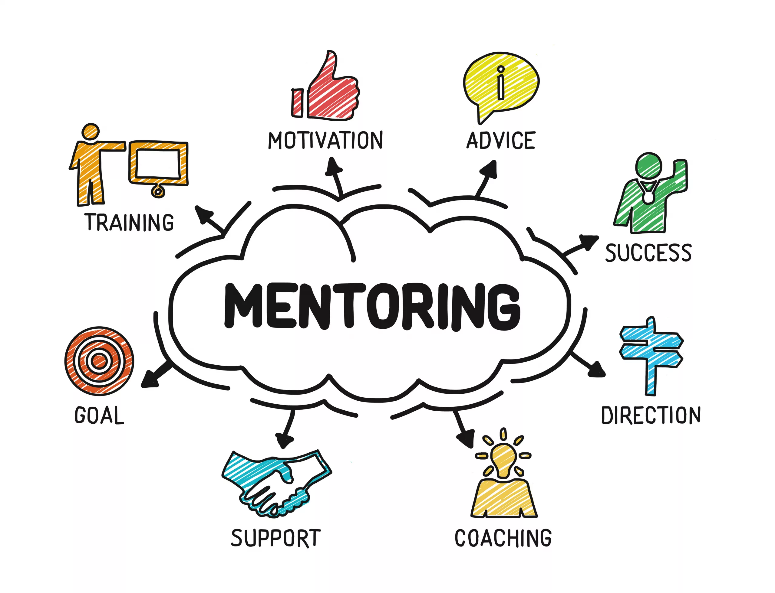
What does mentoring mean to you? Image shows people trying to climb up a series of blocks, receiving help from each other in the form of hands up and ladders.
We as Paramedics do have the benefit of information and support available from unions and the College of Paramedics, but we don’t have an easy to access resource of helpful, responsive, qualified mentors who are there to help you:
Progress
Network
Make informed career choices
Make informed course choices to spend your money and time effectively and efficiently
Find guidance (and perhaps inspiration) when you are unsure where to take your career or what is next for you.
Learning and leadership – two interconnected terms relating to mentoring
If you are interested in becoming a mentor for Article 999 please email me at article999uk[at]gmail.com with your name, role (and registration number if you are registered), clinical background and experience, area of specialist interest(s), and mentoring experience and qualifications. Please note this is currently voluntary, but committed mentors will be contacted in the future as this project develops and grows. Mentors will also receive a certificate that could be used for CPD portfolios.
If you are interested in being mentored then please let us know by liking this post, commenting and/or sharing it.
Motivational quote “Leadership is unlocking people’s potential to become better” appearing behind torn blue paper.
“Mentoring is a brain to pick, an ear to listen, and a push in the right direction.”
John C. Crosby (one of many quotes that describe effective mentoring)
This is a discussion I have had with several people recently. I’ve observed the same discussion on social media: Is it better to specialise by taking specific job roles or courses, or to keep up with the ‘jack of all trades’ role of a Paramedic?
Our profession is at a point of developing specialisms that seem to be staying put. We can now work in primary and urgent care, in research, in palliative care, resuscitation and education roles, or in other community roles such as rapid response. There are ACCP routes popping up around the country. There are resuscitation roles in specialist hospitals, and community roles that focus expertise on specific conditions. I can see a potential future for new staff being one of a few years of general practice, followed by a series of options containing different specialities and of course, different rotas. This is the way other professions have gone and of course, Paramedicine is young compared to the role of a Doctor or Nurse. Perhaps it is only natural for career paths like these to develop over time. It certainly makes for a more interesting, unique career, and it allows us to develop expertise. Specialising also contributes to a clear CV that should help lead to further roles in the area. It could be argued that without emphasising those areas of expertise on our CV, we might stand out less to a potential employer.
Some argue that remaining general means we never develop expertise in any given area, hence the ‘jack of all trades’ expression that I have heard previously expressed about our profession – we all know how that expression ends. Can anyone be a master of all?
Many of the roles currently available offer part-time work. I am working in one of these, four days a week. It means the majority of my time is spent with adults, but as I maintain frontline shifts I could still come across paediatrics and maternity jobs. I may specialise in one or two areas, but if this means I do not develop in other areas then I am not being honest with myself: I need more CPD related to those areas I now come across less because I am at risk of deskilling. If I want to develop as a Paramedic then surely I need to develop in all areas of practice.
My answer to this conundrum is simple: If you do not intend to maintain any frontline work, you do not need to remain general. But if you intend to keep up any of that work, even if this will only be occasional, you must develop in all areas within your remit. You may still specialise and opt for higher level courses in the areas that take up the majority of your time, but it seems sensible to ensure you develop in all the areas you work in. It’s those areas we see the least that will make us the most hesitant. I cannot safely leave my paediatric knowledge behind with what I learned several years ago when I could still see young patients. I cannot stop learning about maternity when those jobs have the potential to be so time-critical. I don’t want to stop focusing on trauma.
I believe to be the best for our patients, we must juggle all areas. Our expertise is then not necessarily about the courses we have done but the experience we have gained. As I spend the majority of my time working in one or two areas, I could argue those are my specialisms – but I am also still remaining general.
There are so many different recommendations of ECG Interpretation. Some books have 5 steps, some 6, some 9, some 11. Below, I have listed a few common ones in the hope of highlighting the differences – and the similarities. I have tried to focus on rhythm strip analysis only but some methods combine the rhythm strip with the 12 lead analysis. I have only included those that are publicly available, so although some fantastic methods are taught on courses, I can’t reference them and have subsequently omitted them. However, if you have a preference or believe one method should be included below, add it in the comments and it may be featured in a future update.
Method one:
From: Resuscitation Council, Advanced Life Support (2021: 106):
The 6 Steps of rhythm strip analysis:
1. Is there any electrical activity? (Or/and check a pulse)
2. What is the ventricular (QRS) rate?
3. Is the QRS rhythm regular or irregular?
4. Is the QRS complex width normal (‘narrow’) or broad?
— 5. Is atrial activity present?
6. Is atrial activity related to ventricular activity and, if so, how?
Method Two:
From: Charles L. Till, Clinical ECGs in Paramedic Practice (2021: 16):
9 Steps – initially look at lead II, then look at every ECG lead:
ECG Steps 1. What is the rate and rhythm? 2. Are there any P waves and what is their relationship with the QRS complex? 3. What is the duration and morphology of the QRS complex?
4. Is the ST segment isoelectric, depressed or elevated? 5. Are the QT intervals and T waves normal?
Clinical Steps 6. Is the heart generating a palpable pulse of appropriate rate and providing adequate perfusion? 7. Is the rhythm unstable and at risk of deterioration? 8. Does the presenting rhythm support or change your working diagnosis? 9. Are any clinical interventions required?
Method Three:
From: Paul Murray, East of England Ambulance Service, Version 1.0, ECG Recognition – Quick Reference Guide (Feb 2014: 1)
6 steps:
1. ‘QRS Rate’
2. ‘Is the QRS rhythm regular or irregular?’ (and regularly irregular or irregularly irregular?)
3. ‘QRS duration’
4. ‘Are P waves present?’
5. ‘Relationship between P waves and QRS – is there a P wave for each QRS and a QRS for each P wave?’
6. ‘Is the PR interval within normal limits 120-200ms and is it constant?’
(This is basically the same as the RC council method, with one added step; the RC method has one step at the start – a reminder to ensure the leads are connected properly and the patient is alive).
Method Four
From: Fred Kusumoto & Pam Bernath, ECG Interpretation for Everyone – An On-The-Spot Guide (2012: 33-36):
‘Assess the patient: symptoms and physical examination. Appearance, vital signs, physical examination’ -> 1. Is there a P wave in front of every QRS and is the rate between 50-100 beats per minute? 2. Are the ST segments isoelectric?
Each answer leads you to a different figure – an option to confirm you have a normal ECG, evaluation of ST segments, evaluation of arrhythmias, or confirmation time-critical patients.
The normal ECG should have: 1. Rate should be between 50 and 100bpm
2. A P before every QRS (Positive P in lead II).
3. The QRS in V1 should be narrow an negative i.e. an Rs
4. The ST segment should be isoelectric
5. The T wave should be the same direction as the QRS
Method Five:
From: Kuhn, Lang, and Wiesbauer, ECG Mastery: The Simplest Way to Learn the ECG (2014: 141-142)
Summarised – 11 steps (taking you from rhythm analysis into 12 lead analysis. Steps included to highlight how some of the steps above are readdressed or appear later altogether here; other points may appear in front): 1. Rhythm. Criteria for sinus rhythm:
Often basic ECG teaching relies on teaching pattern recognition, which can work well, however when presented with an ECG that isn’t similar to a pattern you have seen, you can run in to problems.
This article will explore two simple concepts that will hopefully help with your ECG reading and understanding of the underlying physiology:
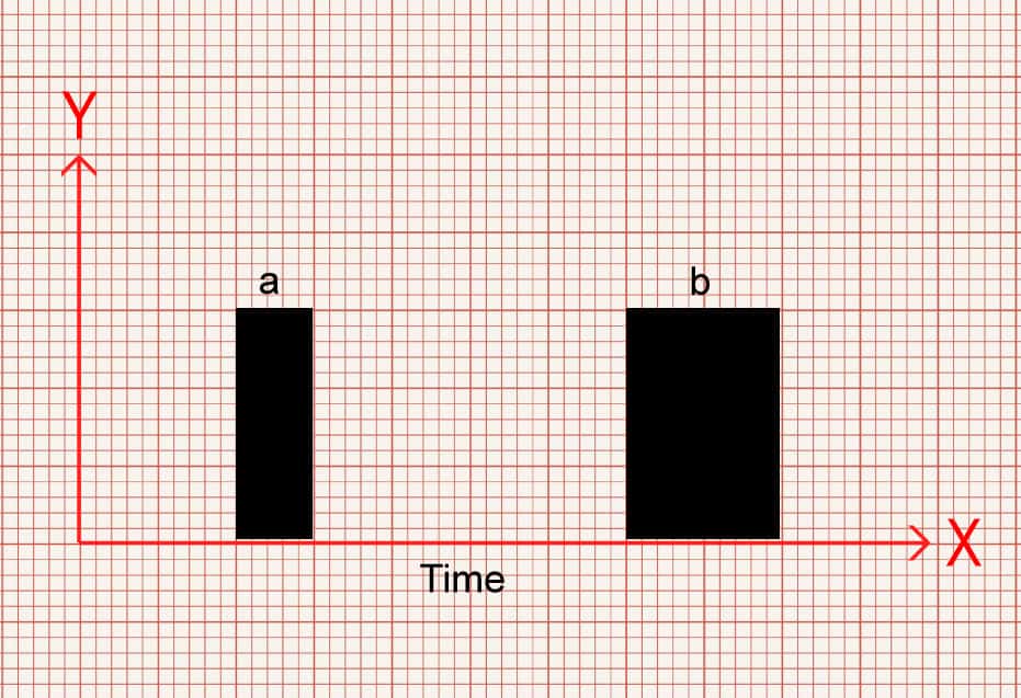
It can be good to think of an ECG as a graph with an X and Y axis. X, horizontally along the bottom represents time. We’ll come to the Y axis later.
Now, we know that X is time, each small square is 40ms (0.04seconds), each large square is 200ms (0.2 seconds), 30 large squares is 6 seconds and so on. That’s reasonably straightforward, but how does it relate to physiology?
Let’s imagine that graph again. And let’s imagine some boxes drawn on. Don’t worry about the Y axis value for now.
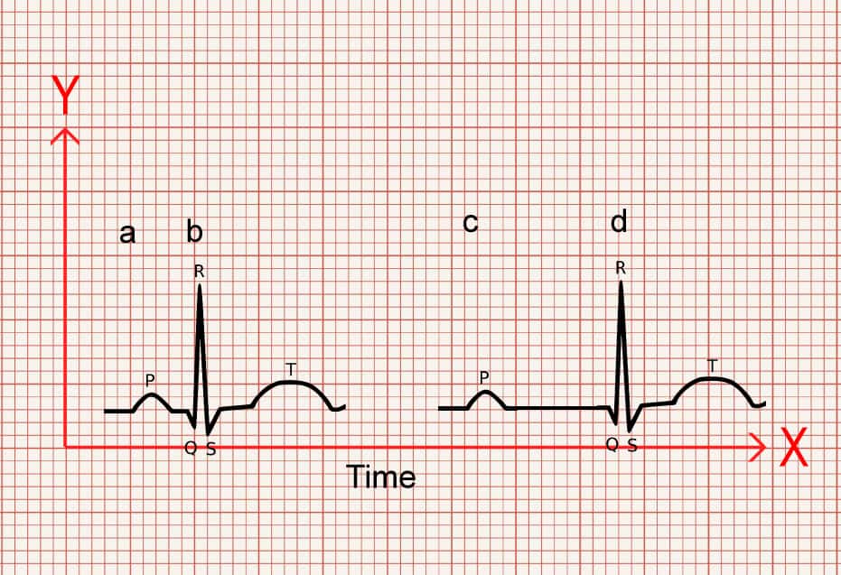
In image 1 you can see a narrow box (a) and a wider box (b). Which one would you say takes up more time? Which one takes up less time?
Box b takes up more of space along the X axis so therefore takes up more time. Box a covers less space on the X axis so takes less time.
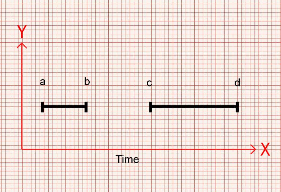
Similarly, we can measure between two points. Look at Image 2. You can see two horizontal lines of different lengths. The gap between a – b is shorter than c -d. So again, which takes up more time?
Gap c-d is wider, takes up more of the X axis and therefore takes up more time.
Stay with me.
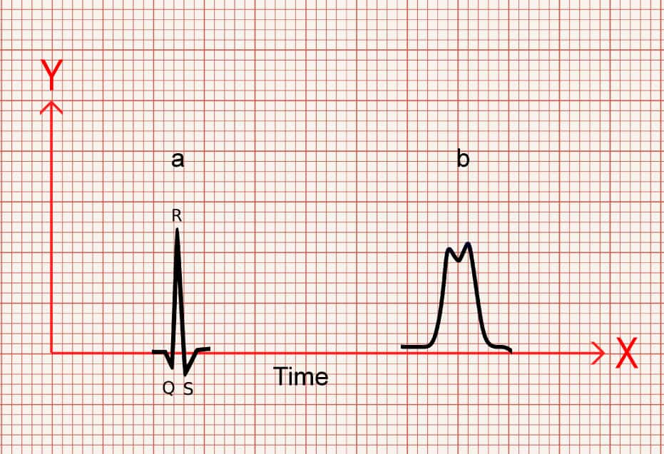
Now, let’s replace those boxes with QRS complexes.
In image 3 there is a narrow QRS (a) and a wider QRS (b). Which one takes up more time?
B is taking up more of the X axis so takes more time.
And again, once more, let’s look at some gaps between two points, but let’s show it as the gap between the P wave and QRS. Which one takes up more time and which takes up less time?
The gap between a-b is shorter than c-d.
So c-d takes up more time.
Probably not ground breaking.
But, what’s another way of thinking about the time something takes? If something takes up more time, you could also say that it is slower, and if something takes up less time, its faster.
Now, remember that an ECG is dealing with electrical conduction.
If on an ECG something is taking up more space along the X axis, therefore taking more time, and is therefore slower, then it makes sense that there is a problem effecting its ability to conduct!
And poor conduction is the underlying issue that what we see in AV blocks and bundle branch blocks.
So if you see a QRS or PR that is wider than normal values, think, something is causing it to be slower and that there is a conduction fault.
Equally if you see a PR interval that is narrower than it is meant to be, taking up less space along the x axis and therefore less time, think, it’s conducting faster than it should and it would alert you to pre-excitation problems, such as accessory pathways in Wolff-Parkinson-White syndrome (WPW effects both the PR interval making it shorter, and has a wider QRS).
Direction
Now, let’s talk about direction.
We know by looking at the ECG paper that vertically, the small boxes, measured in mm, represent millivolts (mV) – a measure of force (sort of). Each 1mm = 0.1mV, 10mm = 1mV. This also means that it has what’s known as magnitude – or size.
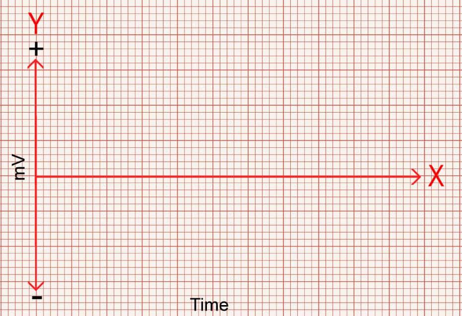
Now, let’s look at the graph again. We know the X axis is time, as discussed above. However, in an ECG the X axis is not simply the bottom, but instead the middle of the graph, with the Y axis extending both positively above the X, and negatively below the X axis.
The X axis can have readings above or below it. This is the isoelectric line.
ECGs pick up electrical charge moving across the heart. And it does so by placing electrodes at different locations around the heart.
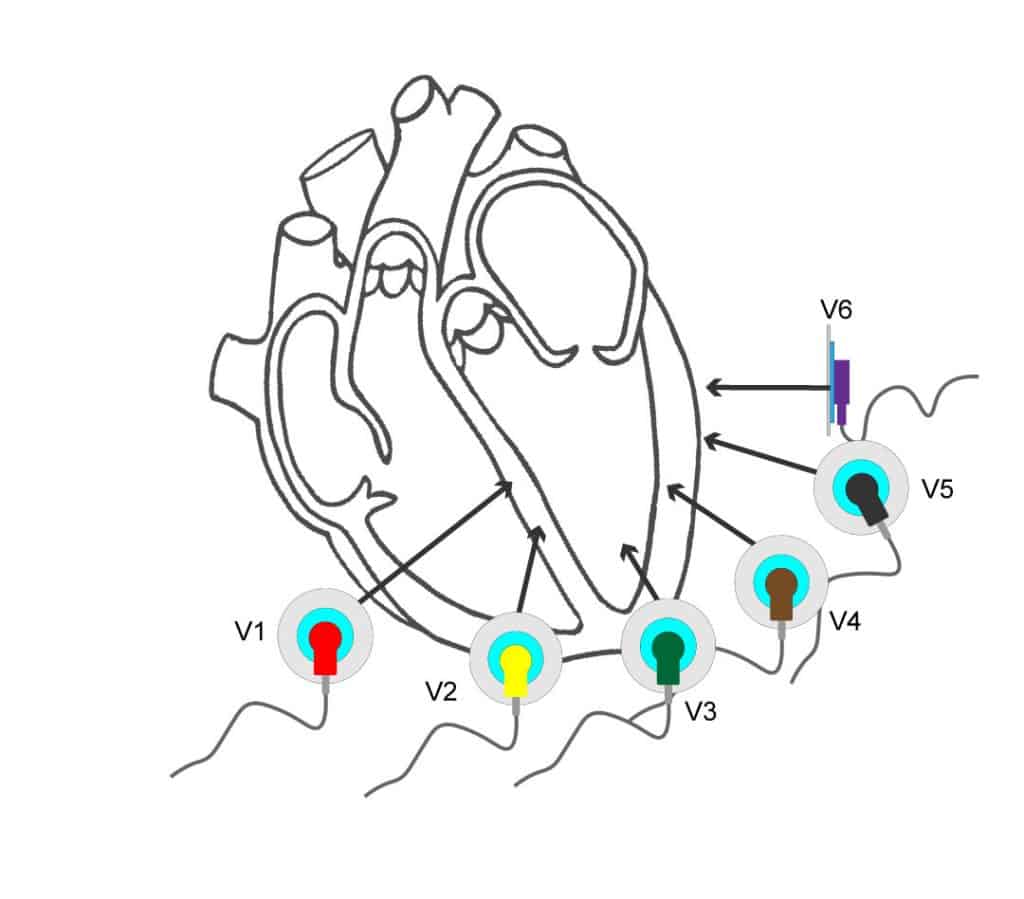
As shown in the diagram below, the electrodes create a view of the heart from different angles, and each view is called a lead. 12 leads (views), but only 10 wires.
The chest leads look at the heart across the transverse plane, that is from the front of the chest to the back, whereas the limb leads look at the heart in the frontal plane, which are essentially from the sides, top and bottom.
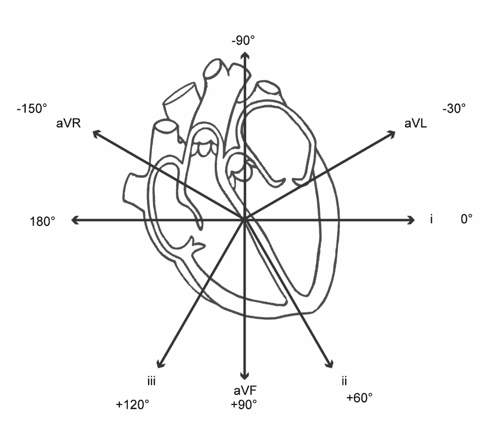
The angles the leads look at the heart are measured in degrees, with lead i being the reference point of 0°.
This is something that is worth learning so that you are able to visualise where each lead “looks”.
Roughly speaking, the limbs leads create the following views of the heart.
avR – Looks from the Right shoulder to the left hip
aVL – Looks from Left shoulder to right hip
i – Looks from the left mid axilla similar to V6
ii – Looks from the left hip to right shoulder
aVF – Looks from the Feet up to the head
iii – Looks from the right hip to the left shoulder
The chest leads create views from the chest wall through to the back
V1 + V2 – intraventricular septum
V3 – V4 – anterior wall of the left ventricle
V5 – V6 – lateral wall of the left ventricle
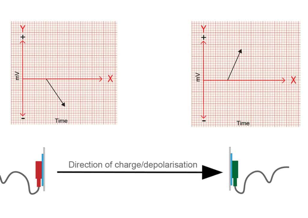
Very simply put, as an electrical charge moves towards an electrode position, this creates a positive reading. As it moves away it creates a negative reading.
Electrodes that are opposite to each other, 180 degrees apart, will be mirrored. One will have charge moving towards it, creating a positive deflection and the opposite side will show a negative deflection as the charge moves away from it.
Leads at 180° from each other.
Slightly more complicated is what happens when the electrical charge isn’t moving directly to, or away from the electrode, but when it is moving at more or less than 180 degrees to the electrodes position.
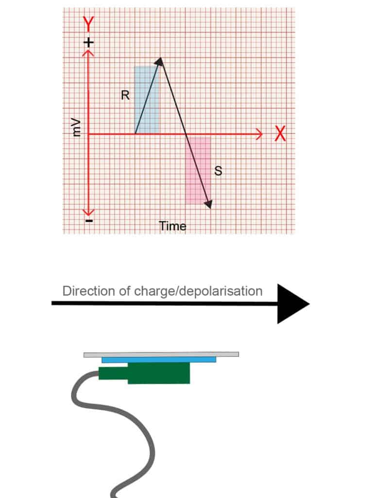
Let’s look at what happens when it moves at 90 degrees to the electrode first.
When the charge is moving at 90 degrees, or perpendicular, to the position of the electrode, it has both a period of positivity and a period of negativity, as the charge moves towards and then away from the electrodes position as it passes by. This is represented on an ECG as an R wave followed by an S wave of equal magnitude.
Quick tip – an R wave is any positive deflection you see, and an S wave is a negative deflection following an R wave BUT it must cross the isoelectric line to be considered negative. The segment from the top of the R wave back to the isoelectric line is not the S wave. The S wave is only the portion below the isoelectric line.
Hopefully this is starting show the relationship between the direction of the charge and the position of the electrode.
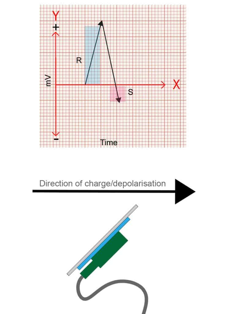
So, what happens if the charge moves at 45 degrees to an electrode’s position? Well, it’s a bit more towards the electrode than 90 degrees, but not straight on. So the tracing will show a predominately positive wave, with a smaller S wave. The opposite will be true if travelling at say 135 degrees away from an electrode.
Hopefully you can see then, that by varying the direction the charge travels, you’ll get different combinations of positive and negative deflections, and that it is relative to the position of each lead/view!
This means that the waves on an ECG not only have a value in magnitude (mV), but also direction. This combination of magnitude and direction is known as a vector.
So not only can the ECG waves tell you the charge in volts, but it can also show you the direction that charge is moving relative to the electrode placement.
Why is this important?
Well, many of the pathologies we come across effect the direction the charge moves across the heart.
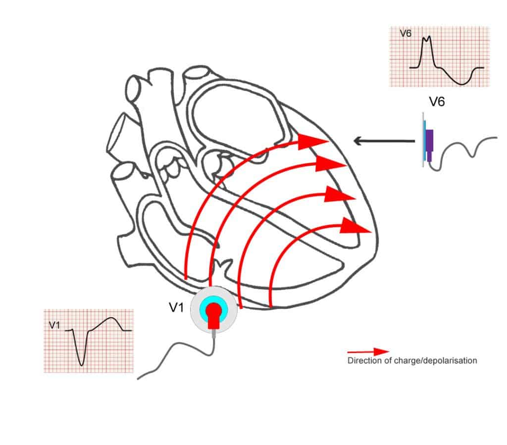
For example, in left bundle branch block (LBBB), the left conduction pathway is faulty. This means all of the electrical charge must come from the right side of the heart, and travels across the heart from right to left through the slower conducting muscle tissue.
V1 looks at the right side of the heart, so, with right to left movement, the charge is moving , slowly, away from V1, giving the classic negative, deep S wave seen in LBBB.
Understanding the concepts of vectors is important if you want to understand axis deviation, reciprocal changes in STEMIs, left anterior and left posterior fascicular blocks, T wave inversions and strain patterns, and how to identify bundle branch blocks.
Final Notes
So, to sum up, understanding how time is displayed on the ECG will lead to better recognition of conduction faults. Understanding that the ECG shows both direction and magnitude means you can easily understand how that charge is moving across the heart. Couple this with further reading around the physiology and pathophysiology and your ECG interpretation should greatly improve!
Just as a final caveat: electrophysiology is complicated. This is a basic description of the principles and vectors, but the images are not exact examples. The detailed process of how the electrodes work, how the leads form their views, the physics behind it and so on are far beyond the scope of this article, but hopefully this has given some insight into the applied nature of ECG.
References
The above post is written by an Article999 contributor and contains a mix of original content and explanations based on the below sources. All images are combined from licensed Adobe Stock images and the author’s own work, as with all Article999 content.
Earlier this year I had the pleasure of speaking with a patient with Functional Neurological Disorder (FND). Ailsa reached out to the College of Paramedics after experiencing an unsettling mixture of treatment by healthcare professionals in a variety of settings. She hopes to encourage healthcare clinicians to learn more about her condition, a functional neurological disorder. Our understanding of this group of conditions is currently being reshaped, so I agree it is another area in which we must stay up-to-date.
The publication of this post on Article 999 comes at a timely moment as just two days ago I read here that a study published in Epilepsy and Behaviour has documented MRI changes in patients with functional seizures. This furthers the point that what science and medicine thought it knew about this – and perhaps other conditions – while not fiction, is also not yet fact.
Members of the College of Paramedics can read the published article in last month’s issue of Paramedic INSIGHT or online here.
In 1999, Anna Bagenholm survived a cardiac arrest after a prolonged down-time and a temperature of just 13.7c. The circumstances of her survival continue to make international news and to inform and inspire research about target temperature management. There have been very few documented cases of survival from such extreme hypothermia, especially with minimal neurological impact. Consider the statistics on neurological impact from ordinary cardiac arrests, with less down-time, and Anna’s case becomes even more extraordinary. But does her survival mean we should target hypothermia in cardiac arrest patients? Should we target hypothermia in ROSC? Let’s have a look at the case and the research that has followed:
The case
Situation: 29 Year Old Female (Anna Bagenholm) is submerged head-first in a hole in the ice in a mostly frozen stream for 80 minutes.
Background: She has no medical history. She has slid down an icy slope while skiing with colleagues.
Assessment: She was conscious for the first 40 minutes, but has been unconscious for 40 mins since. After recovery, she is in cardiac arrest. Her ECG shows asystole. During the resuscitation attempt, the following is also learned:
Temp: 13.7
Pupils: dilated
She is warmed and the resuscitation team are watching an echo of her heart, which begins to move. ROSC was not gained until 4 ½ hours after she fell, and she spent 35 days on life support.
Questions: What would your expectations be of her ROSC management? What would your expectations of her survival and neurological impact be?
Recommendation — today:
Maintain a target temperature at a constant value between 32°C and 36°C for at least 24 h.
Avoid fever (> 37.7°C) for at least 72 h after ROSC in patients who remain in coma.
Do not use pre-hospital intravenous cold fluids to initiate hypothermia.
(Resuscitation Council, 2021)
Decision/Outcome: Not only does Anna survive, her long-term neurological impact is minimal. Have a look at the video:
More Questions:
What target temperature does your Trust follow for ROSC management? Do you think it should be lower, or higher, having seen Anna’s case?
Do you think it’s possible to replicate the results of Anna’s treatment? What makes her situation so different to a cardiac arrest patient who has been treated with target temperature management?
The Research
Have a look at the results of some recent studies into target temperature management, below:
Year
In or out of hospital study
Presenting rhythm
Targets
Outcome
2002
OHCA
Shockable
33 and 37
Positive for survival and neurological impact
2010
OHCA
Shockable
Cooled IV fluids (No target temperature listed)
No difference
2013
OHCA
Both shockable and non-shockable, but presumed cardiac cause
33 and 36
No difference
2014
OHCA
Both shockable and non-shockable
2L of normal saline at 4 degrees, vs standard care
No difference
2018
OHCA
Shockable
32, 33, 34
No difference
2019
OHCA and IHCA
Non-shockable
33 vs 37.5
Improved survival and good neurological outcome (CPC = 1 or 2)
2021
OHCA
Presumed cardiac cause
33 vs 37.5
No difference
Adapted from: Rasmussen and Girotra, 2021.
Final discussion point:
Why can’t controlled studies replicate the kind of outcome that Anna had? Share your thoughts below, or on our Facebook or Twitter pages.
References (excluding videos and direct links above):
Rasmussen, T. P. and Girotra, S. 2021. A Contemporary Update on Targeted Temperature Management, Available at: https://www.acc.org/latest-in-cardiology/articles/2021/11/09/13/16/a-contemporary-update-on-targeted-temperature-management (Accessed 15/05/2022).
Resuscitation Council, 2021. Post-Resuscitation Care Guidelines, Available at: https://www.resus.org.uk/library/2021-resuscitation-guidelines/post-resuscitation-care-guidelines (Accessed 15/05/2022).