Category: Circulation

  • Could This Be The Next Tourniquet?

    Could This Be The Next Tourniquet?

    “At Pentland Medical, we are proud to introduce innovative new products to the UK healthcare system. Hemashock not only has the potential to save lives but improve the longer term outlook for survivors of cardiac arrest and severe shock. We are excited to be part of this journey and show that there is a better way which will lead to better outcomes for patients” – Jamie Munro, Pentland Medical

    A new product in the UK market, the Hemashock from Pentland Medical, acts as an ‘autotransfusion tourniquet’ in cardiac arrest and hypovolemic shock. Rather than merely working as a tourniquet, these auto-transfusion tourniquets ‘sends blood from the legs to the core’. Here’s more about how this specific product works:

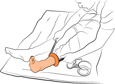
    And here’s how it could look in practice for a solo-rescuer application (graphic only):
    There is some limited evidence of the positive impact of this device. In one case of a PPH:

    ‘IV fluids and Pitocin 20 mg IV had no effect on bleeding or BP. An auto-transfusion tourniquet (A-TT), (HemaShock, OHK Medical Devices, Tirat Carmel, Israel) was placed on one of her legs. Within 1 minute blood pressure started to increase and heartrate slowed.’

    Link to that article here.

    Another case links survival from a presumed cardiac etiology to the application of this device. This is of course difficult to prove, and there are a number of variables that could have impacted the positive outcome – not just to ROSC (CPR quality, identification and/or treatment of other reversible causes, number of shocks) but also to discharge – in particular, target temperature management was used, something still under study and recently removed from Resuscitation Council guidelines following a lack of consistent evidence for its use. However, the article certainly reads convincingly – a patient who is in refractory VF, who swiftly develops ROSC following application of the device.

    Here’s the link to that article. 

    In fact, in a clinical validation at one medical centre, the device appears to have resulted in a higher chance of ROSC in cardiac arrest patients who have had prolonged resuscitation and poor indicators for survival. Notably though, only one patient survived to discharge.

    For further medical evidence relating to the product, please follow this link to the product page. 

    Of course, application of any new product comes with barriers, and as with most interventions we must ask if it is not only necessary but also if it causes any delay or depreciation in quality of the basic and most effective interventions in a medical arrest. This study investigated the use of auto-transfusion tourniquets during cardiac arrest and found that an effective team can deploy them without impacting CPR quality.

    Limitations:

    There are currently no large-scale clinical trials of this device, and the device does not appear to have been tested independently of other variables e.g. target temperature management in ROSC, which is another experimental area given its addition and subsequent removal from the Resuscitation Council guidelines. Additionally, despite auto transfusion tourniquets being investigated in the study above from a team-working perspective, the actual speed of application does not appear to have been examined, something which has been heavily investigated in ordinary tourniquets. Additional human factor elements should be considered here, too – the minimum staff numbers required for application, and the minimum training required for efficient application are both high-stake areas that require further investigation.

    However, the product remains an intriguing dual-purpose device with demonstrable results, and one that adds a positive vision to the future of cardiac arrest management and hypovolemia.

    Summary:

    Please note, this article has been produced by a request from Innovel Medical. No financial or other incentive beyond mere site promotion has been involved, no practical exposure to the product mentioned has been experienced, and this article does not constitute a product recommendation. As always, Article 999 is disconnected from any employers the author works for (see the full disclaimer via the site menu).
  • If you, like me, have never internalised the whole ‘Hi I See…’/HIS thing for areas of the heart…

    If you, like me, have never internalised the whole ‘Hi I See…’/HIS thing for areas of the heart…

    If you, like me, have never internalised the whole HISS thing for areas of the heart and are fed up with people telling you it’s easy, or to visualise it, STOP trying. Keep it simple. People think this is simple but it is only for them. Instead, think about where you apply the ECG leads. Visualise that.

    V1 and V2 are placed on the sternum. You can think sternum for septal, or ‘high anterior’ (as they are high up on the front of the chest).

    Leads v3 and v4 are placed on the front of the chest. They are anterior.

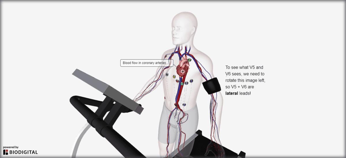
    To see where leads V5 and V6 go, you have to turn an image of the heart to the left. They are lateral leads!

    Try imagining you are the photographer in this 3D image. To see V5 and V6 clearly, you have to be standing on the patient’s left side.

    To remember the limb leads, inFerior is avF, which has two more: ii, and iii. Now all you need to remember is that lateral is I and avL.

    Does it work for you? Let me know. Now to go back and tell my old student paramedic self….

  • Revision Notes: Narrow-Complex Tachycardias – Features

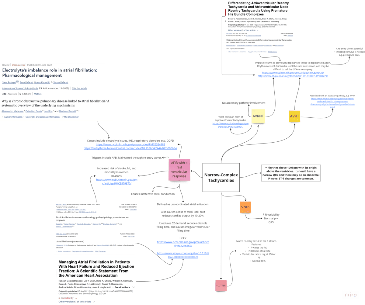
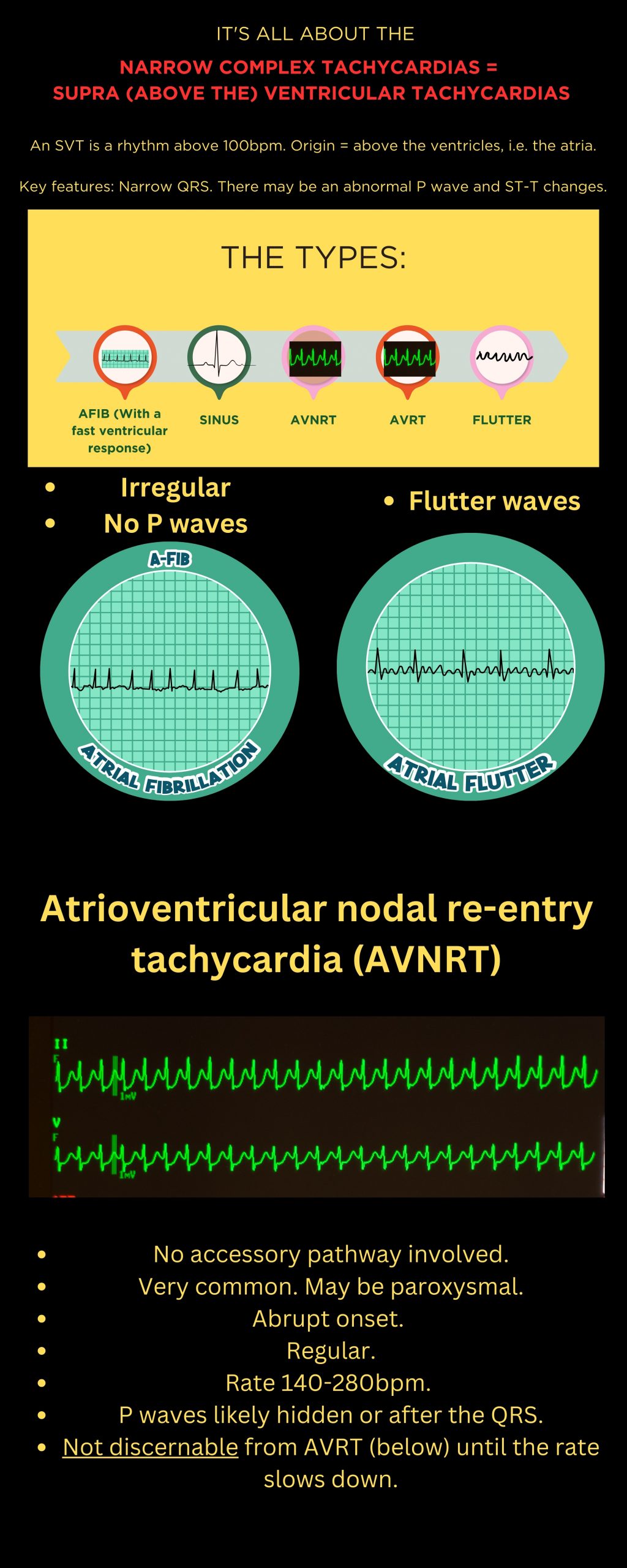
    Revision Notes: Narrow-Complex Tachycardias – Features

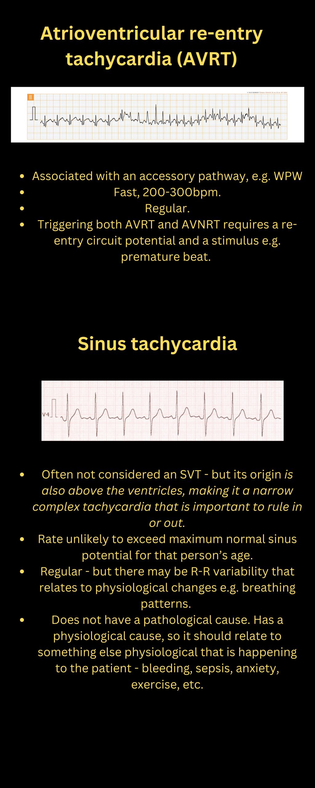
    Have a closer look at the notes in the PDF attachment:

  • The Decompensating Child: Blood Pressure Ranges to Watch

    The Decompensating Child: Blood Pressure Ranges to Watch

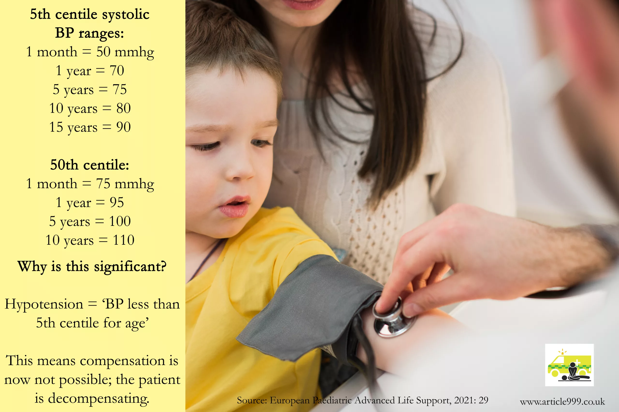
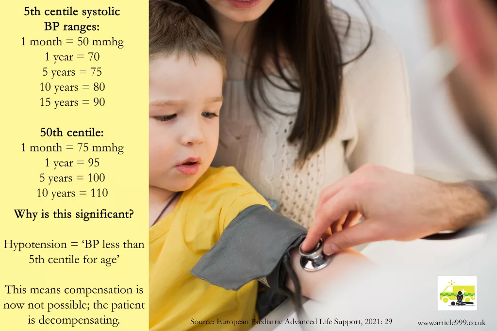
    Image showing the 5th and 50th centile blood pressure ranges for children from 1 month-15 years.
  • Methods of ECG Interpretation

    Methods of ECG Interpretation

    There are so many different recommendations of ECG Interpretation. Some books have 5 steps, some 6, some 9, some 11. Below, I have listed a few common ones in the hope of highlighting the differences – and the similarities. I have tried to focus on rhythm strip analysis only but some methods combine the rhythm strip with the 12 lead analysis. I have only included those that are publicly available, so although some fantastic methods are taught on courses, I can’t reference them and have subsequently omitted them. However, if you have a preference or believe one method should be included below, add it in the comments and it may be featured in a future update.

    Method one:

    From: Resuscitation Council, Advanced Life Support (2021: 106):

    The 6 Steps of rhythm strip analysis:

    1. Is there any electrical activity? (Or/and check a pulse)

    2. What is the ventricular (QRS) rate?

    3. Is the QRS rhythm regular or irregular?

    4. Is the QRS complex width normal (‘narrow’) or broad?


    5. Is atrial activity present?

    6. Is atrial activity related to ventricular activity and, if so, how?

    Method Two:

    From: Charles L. Till, Clinical ECGs in Paramedic Practice (2021: 16):

    9 Steps – initially look at lead II, then look at every ECG lead:


    ECG Steps
    1. What is the rate and rhythm?
    2. Are there any P waves and what is their relationship with the QRS complex?
    3. What is the duration and morphology of the QRS complex?

    4. Is the ST segment isoelectric, depressed or elevated?
    5. Are the QT intervals and T waves normal?
     

    Clinical Steps
    6. Is the heart generating a palpable pulse of appropriate rate and providing adequate perfusion?
    7. Is the rhythm unstable and at risk of deterioration?
    8. Does the presenting rhythm support or change your working diagnosis?
    9. Are any clinical interventions required?

    Method Three:

    From: Paul Murray, East of England Ambulance Service, Version 1.0, ECG Recognition – Quick Reference Guide (Feb 2014: 1)

    6 steps:

    1. ‘QRS Rate’

    2. ‘Is the QRS rhythm regular or irregular?’ (and regularly irregular or irregularly irregular?)

    3. ‘QRS duration’

    4. ‘Are P waves present?’

    5. ‘Relationship between P waves and QRS – is there a P wave for each QRS and a QRS for each P wave?’

    6. ‘Is the PR interval within normal limits 120-200ms and is it constant?’

    (This is basically the same as the RC council method, with one added step; the RC method has one step at the start – a reminder to ensure the leads are connected properly and the patient is alive).

    Method Four

    From: Fred Kusumoto & Pam Bernath, ECG Interpretation for Everyone – An On-The-Spot Guide (2012: 33-36):

    ‘Assess the patient: symptoms and physical examination. Appearance, vital signs, physical examination’ ->
    1. Is there a P wave in front of every QRS and is the rate between 50-100 beats per minute?
    2. Are the ST segments isoelectric?

    Each answer leads you to a different figure – an option to confirm you have a normal ECG, evaluation of ST segments, evaluation of arrhythmias, or confirmation time-critical patients.

    The normal ECG should have:
    1. Rate should be between 50 and 100bpm

    2. A P before every QRS (Positive P in lead II).

    3. The QRS in V1 should be narrow an negative i.e. an Rs

    4. The ST segment should be isoelectric

    5. The T wave should be the same direction as the QRS

    Method Five:

    From: Kuhn, Lang, and Wiesbauer, ECG Mastery: The Simplest Way to Learn the ECG (2014: 141-142)

    Summarised – 11 steps (taking you from rhythm analysis into 12 lead analysis. Steps included to highlight how some of the steps above are readdressed or appear later altogether here; other points may appear in front):
    1. Rhythm. Criteria for sinus rhythm:

    • Are the P waves positive in I and II?
    • Is there a QRS complex after each P wave?
    • Are the PR intervals constant?
    • Are the RR intervals constant?

    Ask: Is it sinus?

    2. Heart rate

    3. P waves – atrial enlargement present?

    4. PR interval

    5. QRS axis

    6. QRS duration

    7. Rotation

    8. QRS amplitude

    9. QRS infarction signs

    10. ST-T segment

    11. QT duration, T-U waves


    That’s it for today, though you may find other methods at useful courses such as Mark Whitbread’s ECG Bootcamp.

    Do you have any recommendations, preferences, or opinions? Add them below!