There are so many different recommendations of ECG Interpretation. Some books have 5 steps, some 6, some 9, some 11. Below, I have listed a few common ones in the hope of highlighting the differences – and the similarities. I have tried to focus on rhythm strip analysis only but some methods combine the rhythm strip with the 12 lead analysis. I have only included those that are publicly available, so although some fantastic methods are taught on courses, I can’t reference them and have subsequently omitted them. However, if you have a preference or believe one method should be included below, add it in the comments and it may be featured in a future update.
Method one:
From: Resuscitation Council, Advanced Life Support (2021: 106):
The 6 Steps of rhythm strip analysis:
1. Is there any electrical activity? (Or/and check a pulse)
2. What is the ventricular (QRS) rate?
3. Is the QRS rhythm regular or irregular?
4. Is the QRS complex width normal (‘narrow’) or broad?
— 5. Is atrial activity present?
6. Is atrial activity related to ventricular activity and, if so, how?
Method Two:
From: Charles L. Till, Clinical ECGs in Paramedic Practice (2021: 16):
9 Steps – initially look at lead II, then look at every ECG lead:
ECG Steps 1. What is the rate and rhythm? 2. Are there any P waves and what is their relationship with the QRS complex? 3. What is the duration and morphology of the QRS complex?
4. Is the ST segment isoelectric, depressed or elevated? 5. Are the QT intervals and T waves normal?
Clinical Steps 6. Is the heart generating a palpable pulse of appropriate rate and providing adequate perfusion? 7. Is the rhythm unstable and at risk of deterioration? 8. Does the presenting rhythm support or change your working diagnosis? 9. Are any clinical interventions required?
Method Three:
From: Paul Murray, East of England Ambulance Service, Version 1.0, ECG Recognition – Quick Reference Guide (Feb 2014: 1)
6 steps:
1. ‘QRS Rate’
2. ‘Is the QRS rhythm regular or irregular?’ (and regularly irregular or irregularly irregular?)
3. ‘QRS duration’
4. ‘Are P waves present?’
5. ‘Relationship between P waves and QRS – is there a P wave for each QRS and a QRS for each P wave?’
6. ‘Is the PR interval within normal limits 120-200ms and is it constant?’
(This is basically the same as the RC council method, with one added step; the RC method has one step at the start – a reminder to ensure the leads are connected properly and the patient is alive).
Method Four
From: Fred Kusumoto & Pam Bernath, ECG Interpretation for Everyone – An On-The-Spot Guide (2012: 33-36):
‘Assess the patient: symptoms and physical examination. Appearance, vital signs, physical examination’ -> 1. Is there a P wave in front of every QRS and is the rate between 50-100 beats per minute? 2. Are the ST segments isoelectric?
Each answer leads you to a different figure – an option to confirm you have a normal ECG, evaluation of ST segments, evaluation of arrhythmias, or confirmation time-critical patients.
The normal ECG should have: 1. Rate should be between 50 and 100bpm
2. A P before every QRS (Positive P in lead II).
3. The QRS in V1 should be narrow an negative i.e. an Rs
4. The ST segment should be isoelectric
5. The T wave should be the same direction as the QRS
Method Five:
From: Kuhn, Lang, and Wiesbauer, ECG Mastery: The Simplest Way to Learn the ECG (2014: 141-142)
Summarised – 11 steps (taking you from rhythm analysis into 12 lead analysis. Steps included to highlight how some of the steps above are readdressed or appear later altogether here; other points may appear in front): 1. Rhythm. Criteria for sinus rhythm:
Fast Fact posts are designed to provide quick and easy ways to revise key topics – view a photo with a quote/fact by simply scrolling through the category. Refresh yourself during those middle-of-the-night breaks when you would have aimlessly scrolled through your phone. There is no need to read a lengthy post to do a little CPD. For more fast facts, head here.
Welcome to a brand new resource containing a searchable collection of charities and companies providing support, information, tools, equipment, food banks and more! Categorised by health condition, circumstance or age group and available for all patients in the UK, we hope you can find useful preventative and supportive help when you need it. This is also available for health-care professionals looking for referral tools and CPD.
The information below has been collated by Article 999’s Founder Louise, along with some help from contributors. Within each category are charities or companies that provide services or equipment that can help you. Please see the ‘date added’ to be certain how up-to-date the information is. Please add comments below, like, and share to let us know what you think and if there are any other charities you would like to see listed here.
This information will be added to. Article 999 is currently not endorsed by any of the charities or companies listed below. If you are a charity and would like to see your posters beside your information, or if you would have any queries, please get in touch by emailing article999uk[at]gmail[dot]com.
HCPs, please do share this with your patients and use the information to enhance your referrals. Some entries below include CPD links for you as well as information for your patients.
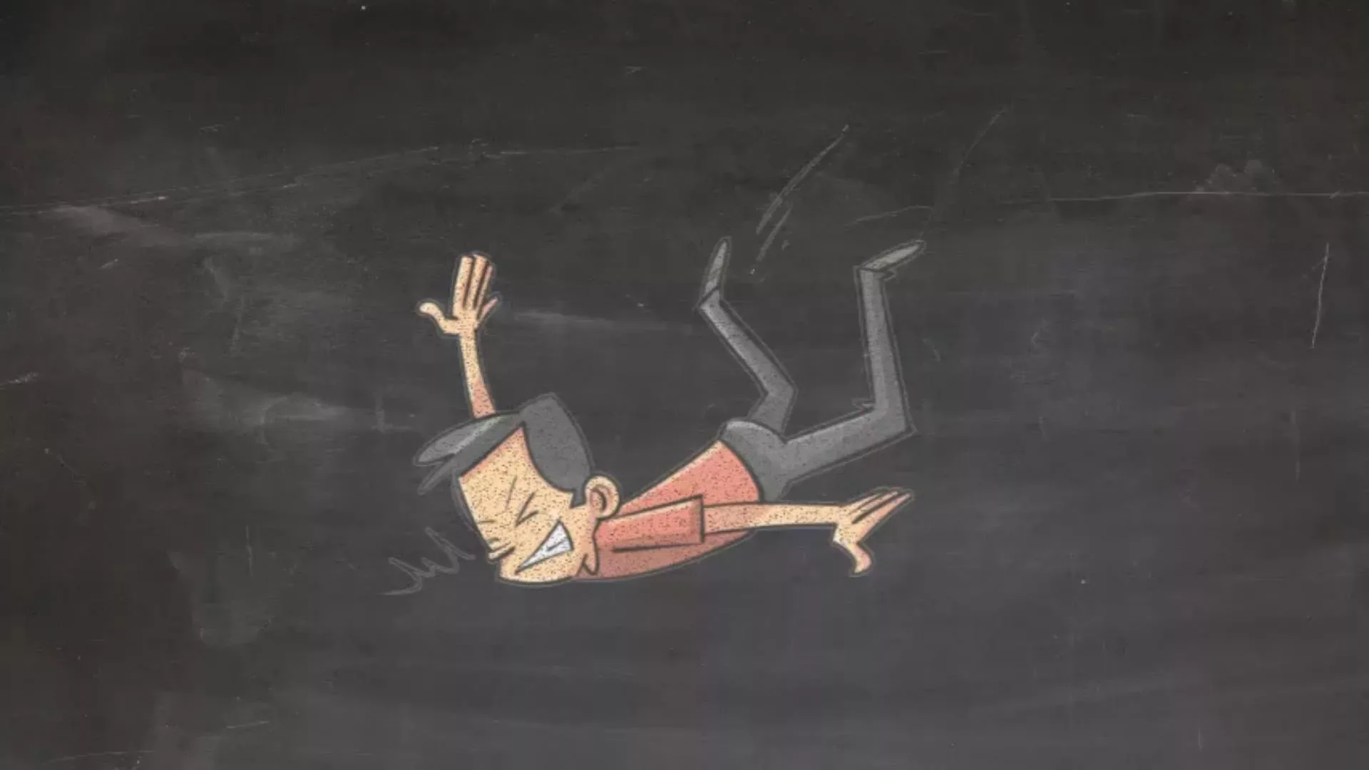
What to Do If You Fall (Adults Under 65) – When to Call for Help
What to do if you fall (adults under 65) and you are not hurt.
More
Adults under 65 who fall regularly may be interested in watching Article 999’s video about when to let your Doctor know about your falls. This is available on the over-65s page but please don’t be offended – the reasons to let your Doctor know will still be applicable.
[tabby title=”For Patients”]
Transcript
*Full references at end of page
If you have fallen over and you’re not sure what to do, or would like some additional information, please keep watching. If an ambulance crew have seen you and you have not attended hospital, this video is also for you.
If you or the person you are watching this for has just fallen and the following apply, please call 999.
If you or they fall again, and the following applies, please call us back:
SYMPTOMS:
You are having difficulty breathing, or have chest pain
You’re unable to move your arms or legs, or they’re numb
You have sudden, new weaknesses
You are dizzy and feel faint when you stand up
You lost consciousness, or have had a seizure, which is not normal for you.
You can’t remember what happened since, or before, you fell over
You have continuous vomiting, or headaches, which is not normal for you.
Your behaviour is different – you feel more confused, or more irritated
call 999
PAIN –
You have neck or back pain
You are unable to get up from the floor
You have another injury that PREVENTS YOU FROM WALKING or causes EXTREME PAIN, for example, you think you might have broken your leg.
call 999
HOW IT HAPPENED –
You have fallen from height – more than 1m
You have landed on your head
call 999
If you have had alcohol, this may be hiding symptoms, making it more difficult for us to assess you. If you are intoxicated, you may be advised to attend a&e to rule out serious complications that are masked by the alcohol.
If you have been advised to attend a&e and have chosen not to, please make sure you have someone who can check in on you over the next 48 hours (NICE, 2014), and ask them to look out for these symptoms.
Also, if you take any medications to thin your blood, AND you have hit your head, you must attend a&e to be monitored and receive a scan of your head, a CT scan. Even if you have not drawn blood, you are still at risk of bleeding internally, especially in the brain because these medications are designed to prevent blood clots.
These medications might be called:
Warfarin
Rivaroxaban
Dabigatran
Apixaban
Edoxaban
Heparin (injection)
You might also be taking any of these antiplatelet medications:
Clopidogrel, aspirin, and others shown on the screen:
Dipyridamole
Prasugrel
Ticagrelor
Cangrelor
(BNF, n.d.)
*These are more concerning when taken in combination, e.g. clopidogrel + aspirin
You should also attend a&e when taking these, or at minimum maintain extreme caution for the same reason. For more information about blood thinners and antiplatelets, please scroll below the video.
What to do if you are on the floor because of a fall, and you’re not hurt
Don’t get up quickly. Roll onto your knees and use nearby stable furniture to push yourself up. Then, sit down until you feel able to continue with your day (NHS, 2021). If you are disabled, ensure use your mobility aids today to prevent yourself from falling again.
Remember, if you have hurt yourself, or you can’t get up, call for help. If you have one, you can press your personal alarm. You can call: family, friends, 999, or 111. Both 999 and 111 will lead to an ambulance arrival with a crew who can assess you and help you up. Both 999 and 111 result in the same ambulance service.
While you wait, the NHS recommends changing your position ‘at least once every half hour or so,’ (NHS, 2021) if you are able to and it is not painful to do so.
Keep yourself warm and let 999 know if anything gets worse.
PREVENTION –
To prevent yourself falling again, remove any objects that might have caused you to fall, avoid drinking too much alcohol, and ensure you wear appropriate safety equipment when performing sports, such as helmets when cycling.
Alter SM, Mazer BA, Solano JJ, et al. (2020). Antiplatelet therapy is associated with a high rate of intracranial hemorrhage in patients with head injuries. Trauma Surgery & Acute Care Open, 5(1), e000520
NICEd. (2014). Head injury: assessment and early management (NICE Clinical Guideline 176). Available at: https://www.nice.org.uk/guidance/cg176 (Accessed 16/06/2020).
Nishijima, D. (2012). Immediate and delayed traumatic intracranial hemorrhage in patients with head trauma and preinjury warfarin or clopidogrel use. Annals of Emergency Medicine, 59(6), pp: 460-468.
North American Thrombosis Forum. (2020). Falls and anticoagulation. Available at: https://natfonline.org/2020/08/falls-and-anticoagulation. (Accessed 18/10/2021).
Probst, M. et al. (2020). ‘Prevalence of Intracranial Injury in Adult Patients With Blunt Head Trauma With and Without Anticoagulant or Antiplatelet Use’. Annals of Emergency Medicine, 75(3), pp: 354-364.
Soliman, Y., Meyer, R., And Baum, N. (2016). Falls in the Elderly Secondary to Urinary Symptoms. Reviews in Urology. 18(1), pp: 28-32.
Stiell, I. et al. (2003). ‘The Canadian C-Spine Rule versus the NEXUS Low-Risk Criteria in Patients with Trauma’, The New England Journal of Medicine. 349. pp: 2510-2518.
Please feel free to share this video with your patients, but please attach this link with it so that all accompanying information is also shared.
For more info about what Article 999 is, and why I am creating videos for patients, please see the ‘About Us’ page and click on the ‘About article 999 for patients’ tab.
Feel free to add comments or feedback. Please let us know how well received this is.
Thank you,
Louise Sopher HCPC Registered Paramedic Article 999 Founder
27:45 additional free support – details. Note: Teams 1:1 support is now a paid service of a one-off fee, set at £15. Support for customisation to ensure the template meets Trust requirements will continue to be offered for free.
Those following Article 999’s Facebook and Twitter pages from December 2022 onwards may have noticed daily posts providing short questions and simple answers, or interesting quotes, on every topic from A-E. This is what Article 999 was originally provided for – short, simple, referenced answers to common questions, and videos to learn and revise practical skills. I hope that by providing these, it’s a little easier to revise common topics, tick off some CPD (remember, you can use my templates for that), and come up with 1-sentence answers when colleagues or patients (or exams) ask you questions. These Q&As are now being archived here, and will steadily begin to be expanded upon. As always, let me know what you think in the comments below.
These ‘often occur due to the confluence of five arterial vessels on the anteroinferior septum’
Reference: Van De Graaff and Rhees, 2011. 256
Beginning with a primary tumour, describe the progression of lung cancer
Primary tumour compresses and invades the surrounding lung tissue
Invasion of chest wall, pericardial cavity, arteries, mediastinum
Metastases into local and distant lymph nodes including the adrenals
Metastases into distant organs
Reference: Bateman and Carr, 2009: 63.
How does capnography work?
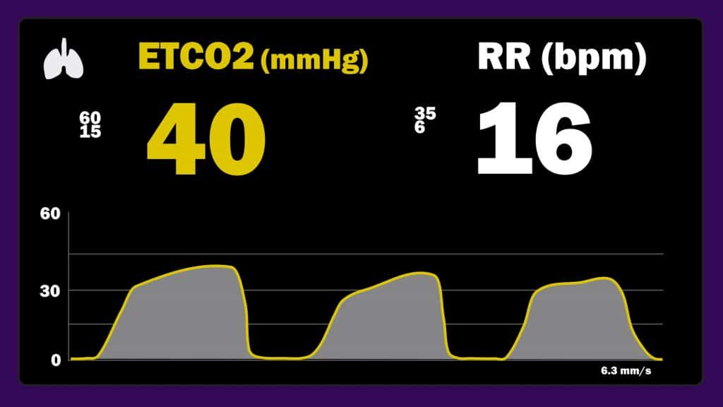
‘Capnography uses infrared light to measure C02 in exhaled air’
Valente, 2010: 1
Image shows ETC02 in mmHg, and a normal ETC02 range is 35-45 mmgHg; the kPa equivalent is 4.7-6.0 (Messina and Patrick, 2022).
Q: Why is adrenaline listed as a treatment for pre-hospital clinicians for asthma, but it’s not part of the RC algorithm?
A: Difficult to answer. Adrenaline is in NO professional guidelines for asthma other than JRCALC. There is limited evidence of benefit compared to selective beta 2 agonists, such as salbutamol, yet may have a worse side effect profile. Anecdotal evidence reports good results in patients who are in extremities or peri arrest
Baggott et al, 2021. https://buff.ly/3X37Nhj
Answered by Adam Philpot
Cardiac
What is the action potential?
The way an electrical impulse ’causes cardiomyocyte cells to depolarise’ and subsequently leads to a ‘chain reaction in adjacent cardiac cells’ so that this impulse moves ‘through the heart’
Reference: Till, 2021:5
Photo: Adobe Stock
How do I interpret the acid-base balance?
This one is a published post – click here to view it.
How do I identify left bundle branch block on an ECG?
All bundle branch blocks will have a widened QRS of >=120ms/3 small squares.
Left bundle, specifically, will also have a negative QRS in V1 and V2 and a positive QRS in V6.
The ECG may also have any of the following features:
an RSR (‘notching’) pattern on the left side of the heart (v5/v6)
a W shape in V1 and an M shape in V6
However, the ECG may not have all of these features, and other features might exist alongside the left bundle branch block that are indicative of the severity of disease – more on this soon.
‘Deficient digestion/absorption’ related to problems with enzymes or ‘an absorptive surface of the GI tract (e.g., inflammatory bowel disease or bacterial overgrowth)
‘Problems with the blood vessels/blood supply (e.g. esophageal varices, mesentric ischemia, GI bleeding)
Abnormalities of the blood supply, which may be directly due to bleeding or result from thrombosis/atherosclerosis
Inflammation including colitis/chron’s disease
Tumours
‘Excess acid secretion’ due to ulcers
Malabsorption in the small intestine
Large intestine obstruction
‘Outpoutchings (diverticula) that prevent the food from going where it is supposed to go’
‘Inability to prevent stomach contents from re-entering the esophagus’ – GERD.
Reference: Berkowitz, 2007: 71
Topic: GU/GI
Q: What is peptic ulceration?
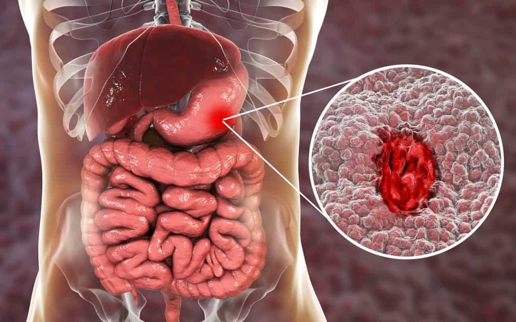
A: An acute or chronic ‘breach in the mucosa (i.e. full thickness loss) of the gastrointestinal tract caused by the action of acidic gastric juice.’
Reference: Bateman and Carr, 2009.
Photo: Adobe Stock
Gastric ulcer. Mucosa of stomach with ulcer and anatomy of human digestive system. 3D illustration
Q: What are the causes of gastritis?
A:
NSAIDS, which ’cause reduced prostaglandin synthesis in the gastric mucosa, leading to exfoliation of surface epithelial cells and inhibition of mucus secretion, thus reducing mucosal defences against acid attack’
Alcohol, which irritates the stomach lining,
Stress, which leads to inflammation
Bile reflux
Bateman & Carr, 2009: 104
Q. What does H.pylori do in the body?
A.
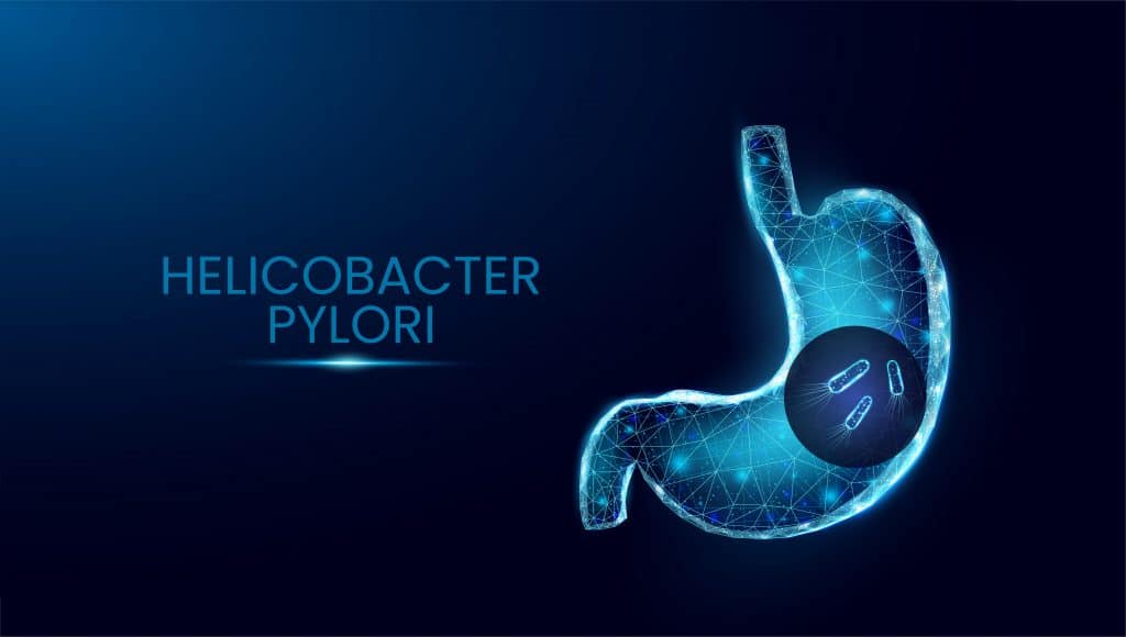
– ‘it lives in the surface mucus of gastric mucosa […] where it secretes a number of toxins that damage surface epithelial cells and break down the mucus barrier’
– It ‘produces a [self-limiting] transient acute gastritis’ and some people cannot ‘clear the organism’, leading to ‘chronic inflammation of the mucosa’
– It can lead to peptic ulceration; it can also produce no symptoms
Ref: Batman & Carr, 2009: 104
Describe the formation of cholesterol stones.
1) The liver secretes excess cholesterol
2) The bile is supersaturated
3) There is now less motility of the gallbladder or/and nucleation (new material forms). Note: reduced motility can also be caused by spinal cord injury or total parental nutrition.
4) Reduced motility leads blocks the release of bile from the liver, which in turn leads to nucleation
5) This leads to stone formation
Bateman & Carr, 2009: 108.
Neuro & Toxins
‘Among the various clinical assessments, the absence of normal sitting balance appears to be a particularly strong predictor of early death; 51% of those with abnormal sitting balance died between 1 and 3 months after the stroke, compared with 11% of patients who were able to maintain an upright sitting position.’
From: R. Anderson, 1992. ‘The Aftermath of Stroke: The experience of patients and their families’, Cambridge University Press: Northamptonshire. p88
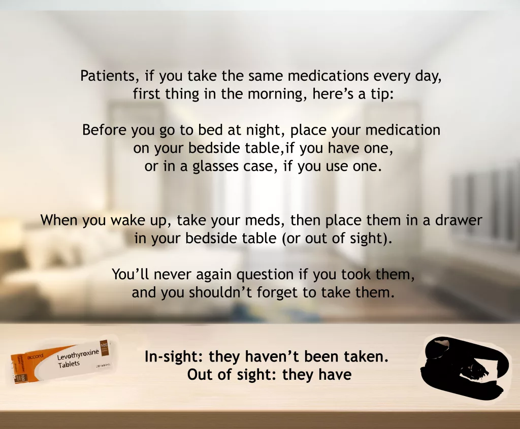
What food interactions are there with levothyroxine?
All drinks containing caffeine – leave at least 30 minutes from taking the tablet before drinking them
Calcium rich foods – the NHS advises waiting 4 hours prior to consumption of these foods
‘a medicine used to lower levels of uric acid’ in the blood and is used to treat gout and kidney stones or, in some cases, for cancer treatment that leads to uric acid accumulation
‘it is not uncommon to “react” to “new” clinical symptoms the same way one reacts to acute injuries. […] For example, an athlete with a stress fracture (a fatigue fracture) in the foot will often state that the symptoms originated during a specific run, perhaps even from a specific step. The injury may accordingly be misclassified as an acute injury. However, the actual cause of the stress fracture is that the specific run was a precipitating event on top of the underlying spectrum of tissue damage on the skeleton from overuse over time. Therefore, these types of injuries should be classified as over-use injuries.’
From: R. Bahr. Ed. 2012. ‘The IOC Manual of Sports Injuries: An Illustrated Guide to the Management of Injuries in Physical Activity’, The International Olympic Committee: Oxford. p2
What are the different grades applied to soft tissue injuries?
1-3/minor, moderate, severe.
Grade 1 ‘will typically damage only a small number of fibres’ and causes ‘pain when the part is used or stressed’; may also lead to ‘swelling and bruising’ but not disability or deformity
2 = ’caused by a more severe mechanism and there will be a larger number of damaged fibres’; there may be ‘a painful loss of function in the muscle’; patients should be referred to physio
3 = ‘complete division of an injured tissue, most commonly ligament, tendon or muscle belly’; ‘complete loss of function in the injured part’; stressing the ‘torn tissue’ does not necessarily cause pain; any deformity or laxity may be present
Purcell, 2003: 41
Q: Define overuse injuries
A: Those that ‘occur gradually’ and ‘exist along a spectrum where the inciting events are below the threshold for clinical symptomatology, but if not rectified, they eventually produce sufficient tissue damage to result in clinical symptoms.’
Ref: Bahr, 2012: 1
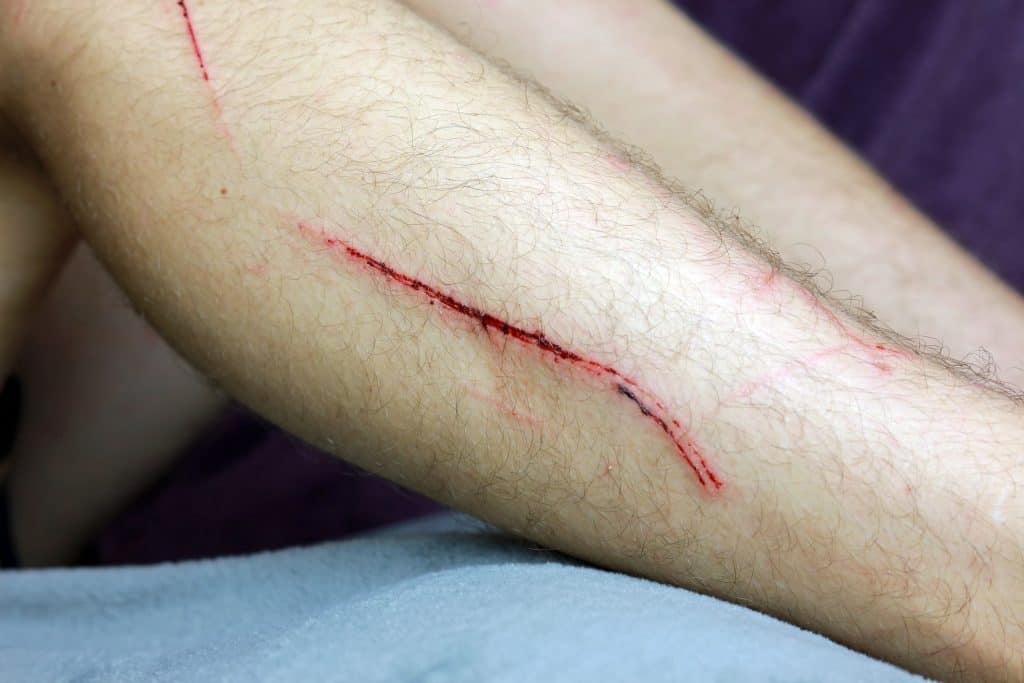
Q. What are the contraindications to the repair of a laceration?
At what age should a child start ‘smiling with meaning’?
5-8 weeks
Reference: Gill and O’Brien, 2007.
What is the definition of a neonate?
‘a newborn infant up to 28 days after their estimated delivery due date’
Reference: Brugha and Marlais, 2013.
Q: How does the size of an infant’s head relate to airway obstruction?
A: Their large heads ‘flex on the neck when the infant is placed in a supine position’, causing ‘potential obstruction of the airway when the conscious level is reduced.’
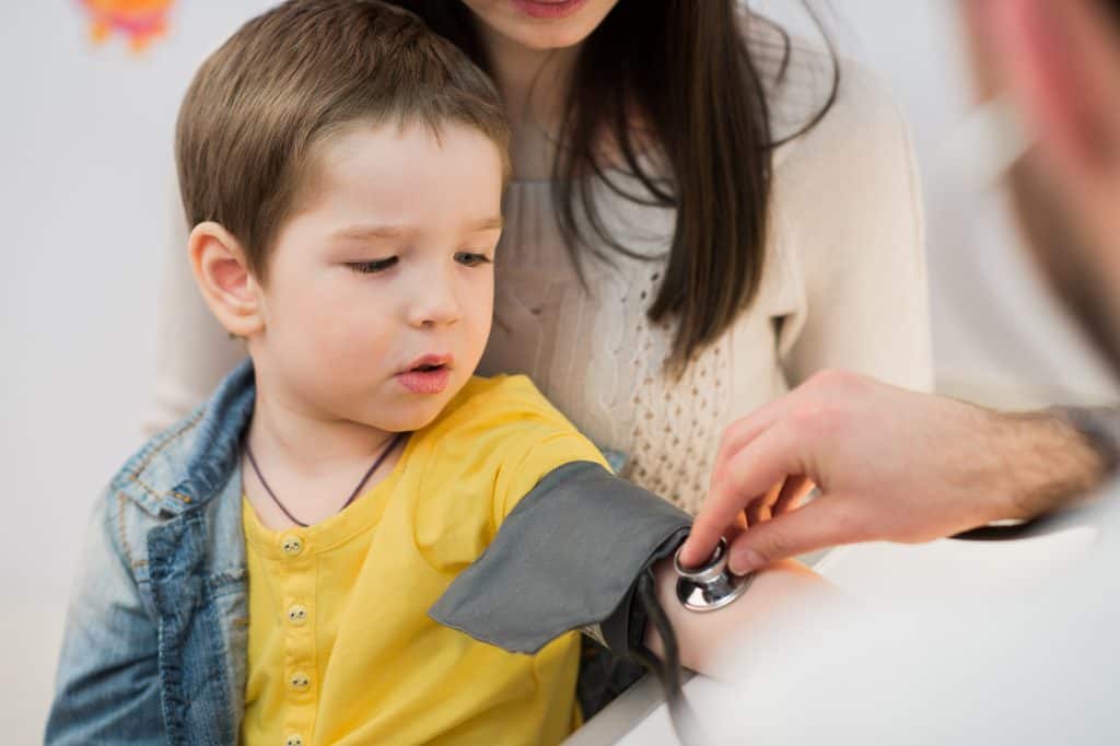
Which is a better indicator of tissue perfusion’ in unwell children and infants – systolic blood pressure or mean arterial blood pressure?
Mean arterial blood pressure – this is an ‘indicator of blood flow’ and better because it takes into account ‘the fact that two thirds of the cardiac cycle is spent in diastole’. Note: not always reliable as patients can have an acceptable mean arterial pressure but poor cardiac output, leading to poor tissue perfusion. The expected mean arterial pressure should be compared to the actual one, found through this formula: ((2x diastolic) + systolic) / 3 = 3 MAP (mmHg)
Resuscitation Council, 2021. European Paediatric Advanced Life Support. 5th ed. London. (p14).
Little boy medical visit – doctor measuring blood pressure of a child.
CPD
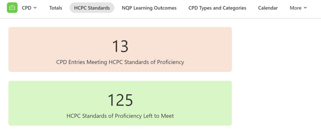
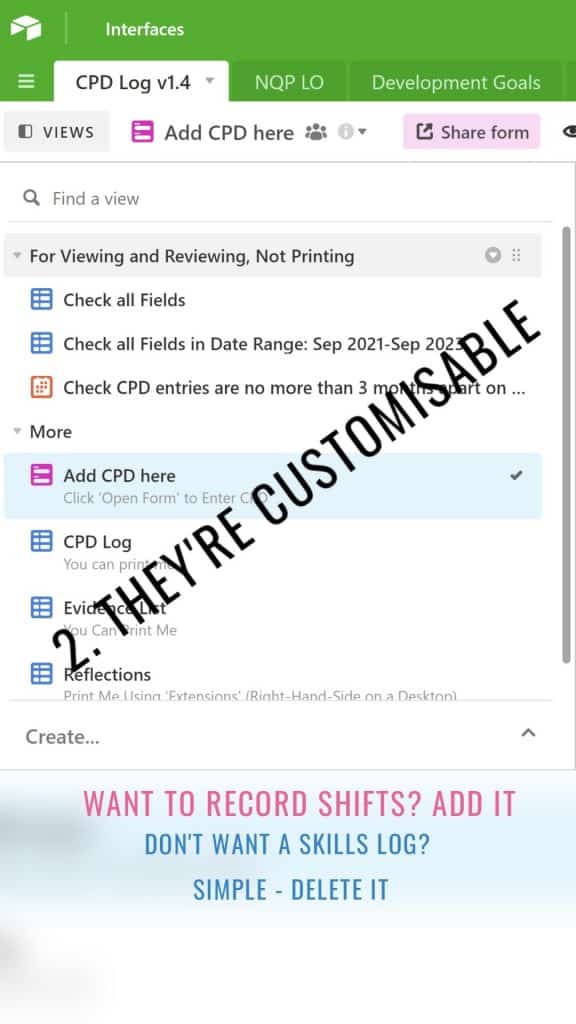
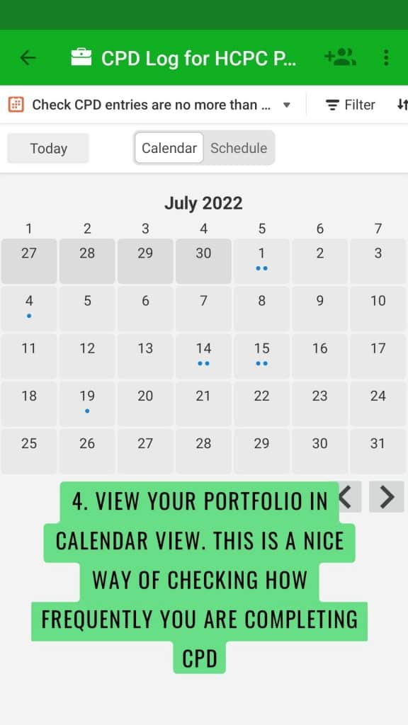
Great things you can do with the CPD templates – visual data demonstrating how many standards you are meeting. This is a free add-on, so feel free to get in touch to ask for help to set it up in your portfolio.
How do I start logging my CPD using Article 999’s templates?
Set up a free Airtable account here. Note: Article 999 is not affiliated with Airtable.
Head to Article 999’s CPD Templates page here. Specifically, click on a) if you are a Paramedic post-NQP, or b) if you are currently an NQP. Don’t worry, both templates are very similar, and if you are in the (b) category, you can continue using the same portfolio when you complete your NQP period.
Click on the ‘use template’ button and add the template to your own account.
Start editing and logging. Click on the ‘Add CPD here’ form, then ‘open form’, save that link everywhere – your phone, your desktop, your notepad, and log away.
Need help? No worries. Email me at article999uk[at]gmail[dot]com
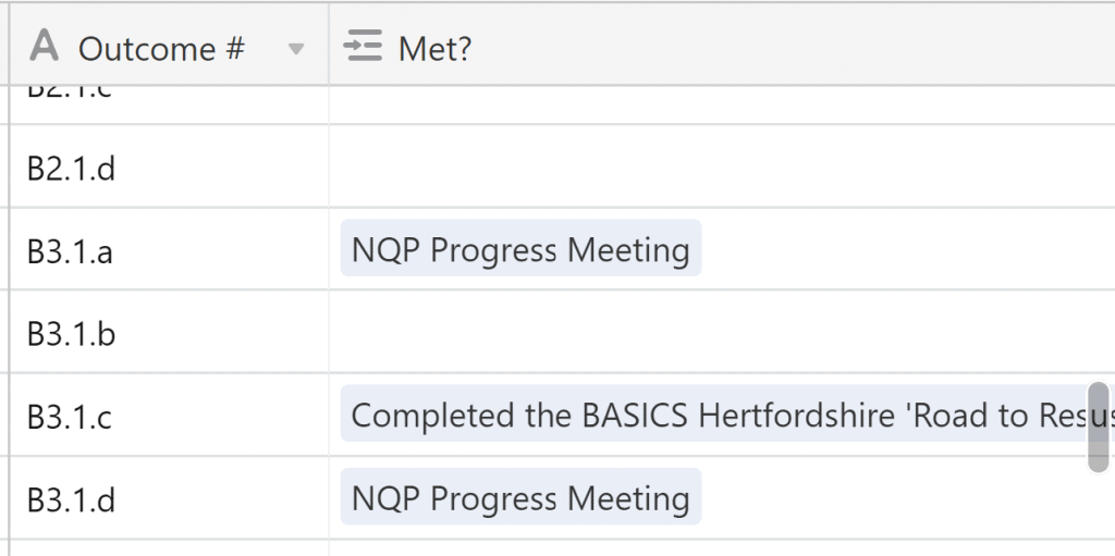
Q: How do I know if I’m meeting the NQP learning outcomes?
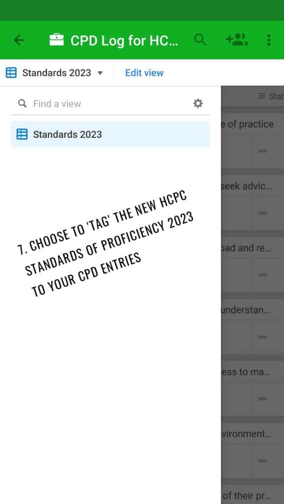
A: All learning outcomes from the NQP portfolio (EEAST version*) are listed in a table. Each time you log your CPD, you can choose to search for a relevant learning outcome, or scroll down the list, then ‘tag’ one to your CPD entry. You can also check how many learning outcomes you have completed by clicking on the specific table and scrolling down (pictured).
*Not endorsed by EEAST but I am a Paramedic working for them.
Here’s a list I’ve had for a while but not published: obvious, less obvious, and utterly surprising reasons your alive patient might be pale. References within. Detailed reference list coming soon.
Image above – a person with vitiligo (Source: Adobe Stock)
Drugs – Amiodarone can cause a ‘bluish-grey’ skin discoloration (Douglas et al, 2013: 44)
Vitiligo (segmental and non-segmental) due to the lack of melanin, causes ‘pale patches of skin’ (Douglas et al, 2013: 46); non-segmental vitiligo ‘is thought to be an autoimmune condition’ (NHS – Vitiligo)