If you’re a UK Paramedic looking for a template to use for your CPD portfolio, here’s a link to one I’ve made. You can download and copy this template into your OneNote or other note application. Simply fill in the blanks, create your own dated list of CPD activities*, and then delete the help info. Keep your completed portfolio available for any HCPC audits, and save the help folder for any questions. The portfolio is referenced and contains direct links with screenshots from useful sources.
*I now also have a free template available for you to use to add your own CPD. Please follow this link for more information. (Feb 2022; link updated Jan 2023)
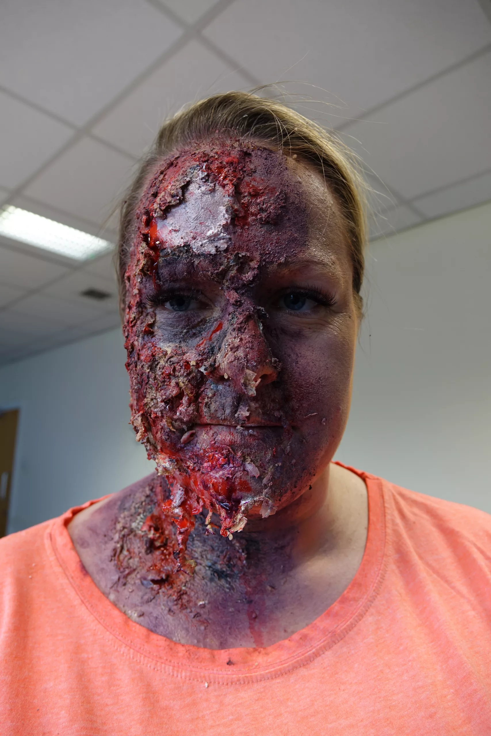
Content warning: This video may contain sensitive content.
This video demonstrates the assessment and management of burns, using equipment that is recommended within the locality at the time of production. Remember to check your local, current guidelines before putting anything into practice. Produced by HCPs and students.
This video demonstrates use of burn gel wraps, which may no longer be used in your ambulance Trust. Please check your local guidelines regarding this. According to the British Burn Association:
Burn gel wraps may be used to provide analgesia, but only after adequate cooling has occurred as they do not actively remove heat from the wound
Varley et al (2014)
Remember to follow good IPC in real life.
The transcript below has been slightly edited, with titles added, to make it easier to read.
You must read the full disclaimer at www.article999.co.uk/disclaimer and check your local guidelines before putting into practice any of our content.
BURNS: The practical stuff
This video contains content some might find sensitive.
Treatment
With all assessments and interventions, use an aseptic technique, especially because ‘burns are prone to infection’
Purcell, 2003: 217
Airway
The patient’s airway might worsen. Here’s why:
‘A burned patient may have a patent airway on the initial evaluation. […] In the time that follows, the face, as well as the airway, will likely swell.’
NAEMT, 2016: 411
… So, consider HEMS for RSI.
Breathing
The patient’s breathing might worsen. Here’s why:
Is there a chest wall burn?
‘Burns constrict the chest wall similarly to having several leather belts progressively tightening around the patient’s chest. As time progresses, the patient cannot move the chest wall to breathe.’
NAEMT, 2016: 412
So, consider HEMS and ventilatory support where needed.
Other considerations:
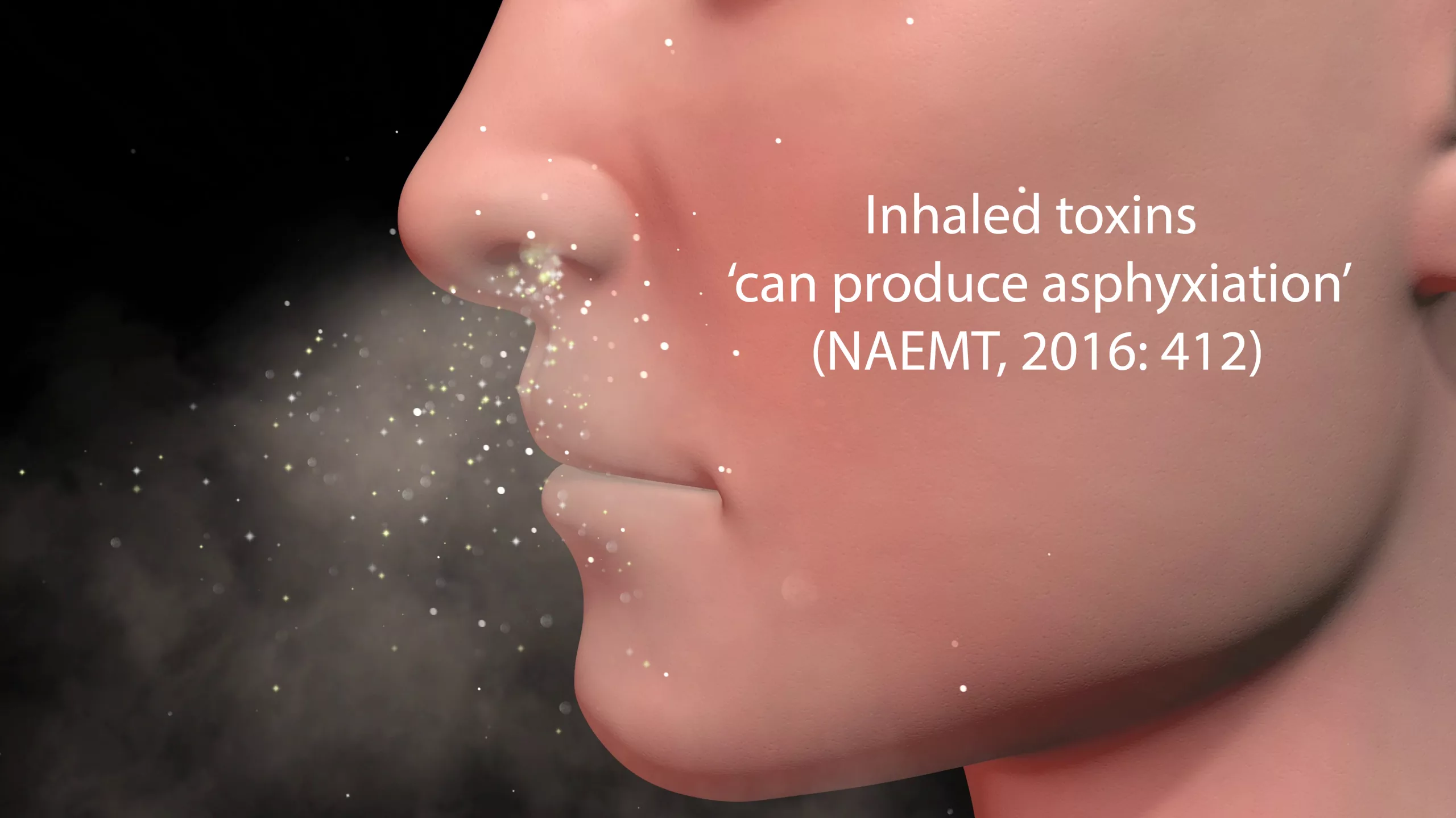
Inhaled toxins ‘can produce asphyxiation’
NAEMT, 2016: 412
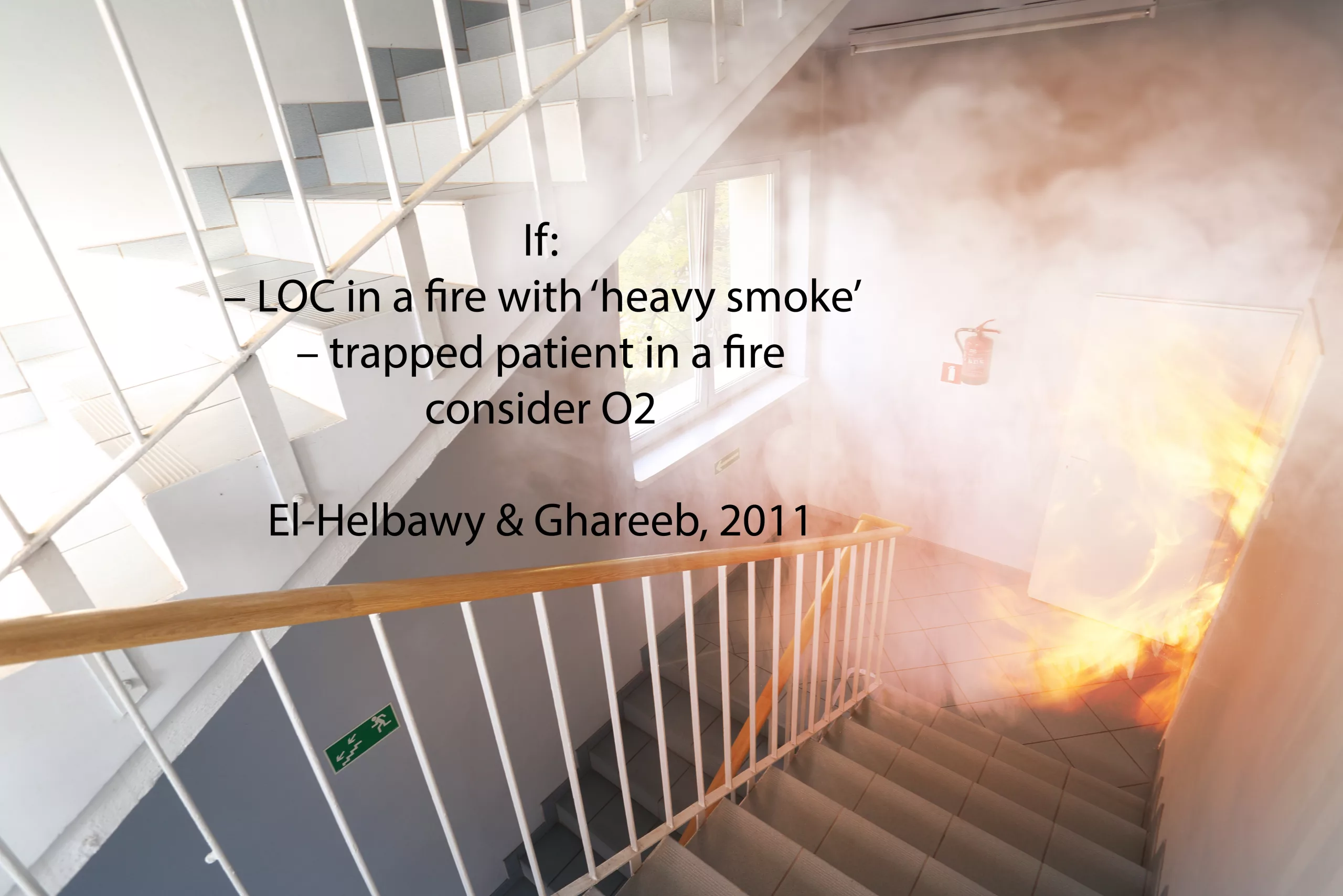
If: – LOC in a fire with ‘heavy smoke’ – trapped patient in a fire consider O2
El-Helbawy & Ghareeb, 2011
Circulation
Swelling might get worse. Here’s what to do about it:
‘Distal limb perfusion may be critically reduced’; ‘Burned extremities should be elevated during transport to reduce the degree of swelling in the affected limb’
NAEMT, 2016: 412
A complication of swelling:
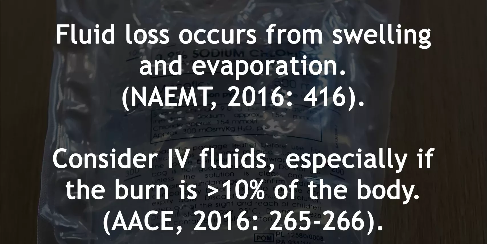
Fluid loss occurs from swelling and evaporation
NAEMT, 2016: 416
Giving Fluids – Pros and Cons
Consider IV fluids, especially if the burn is >10% of the body (AACE, 2016: 265-266). Keep watching for info on how to determine this.
Give fluids with caution. Too much -> ‘cardiac failure, […] infectious complications, acute respiratory distress syndrome, and even death’.
Too little -> ‘hypovolemic shock, organ failure’
(NICE, 2016)
Judicious fluid management of children with severe burn injury can improve the respiratory outcome measures of these children
Duran and Sheridan, 2016
Cannulating – Essentials
When cannulating, ‘placement through the burn is appropriate [only] if no alternative sites are available’
NAEMT, 2016: 412
What about heat?
Patients with burns are not able to maintain their own body heat
NAEMT, 2016: 413
…So, give blankets
ECGs
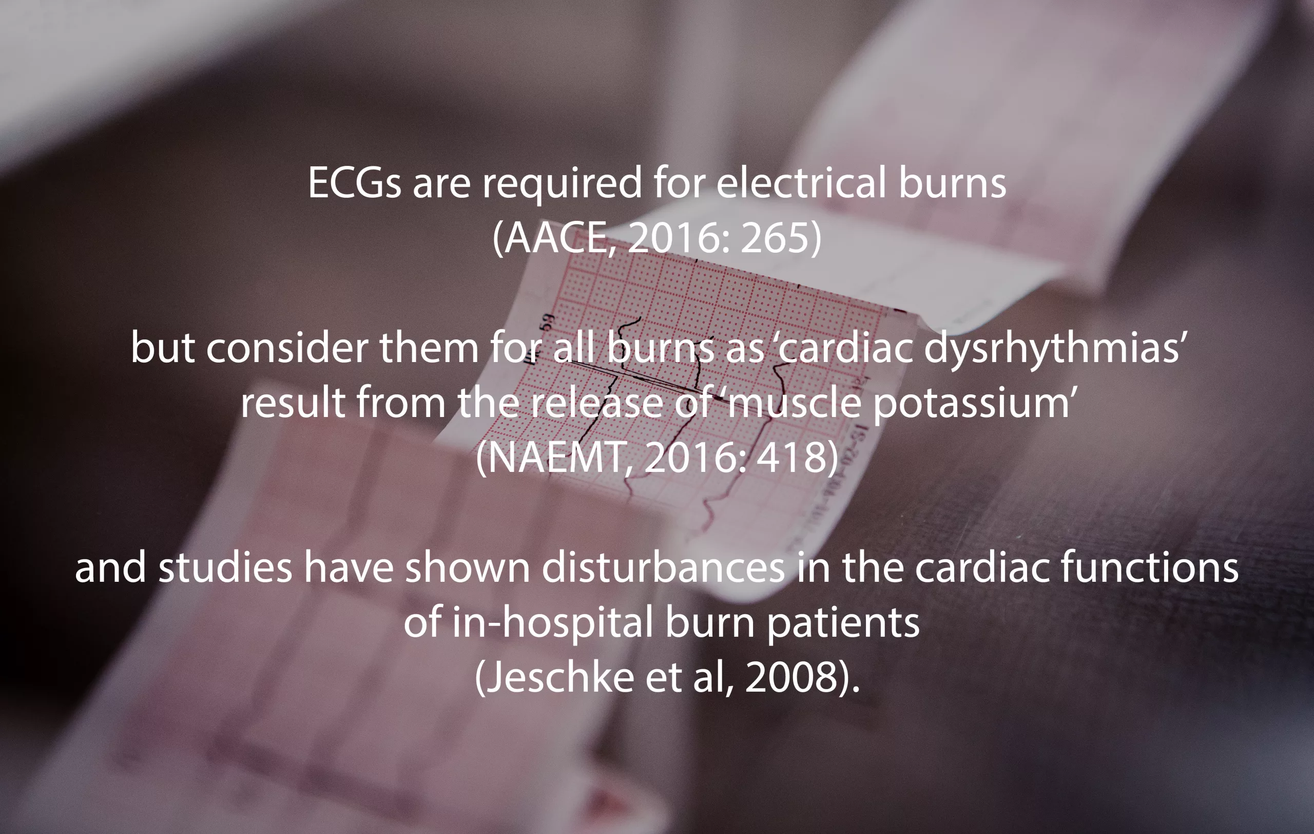
ECGs are required for electrical burns (AACE, 2016: 265) but consider them for all burns as ‘cardiac dysrhythmias’ result from the release of ‘muscle potassium’ (NAEMT, 2016: 418) and studies have shown disturbances in the cardiac functions of in-hospital burn patients (Jeschke et al, 2008).
Disability
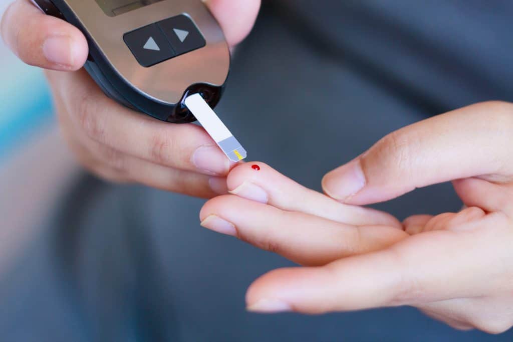
Do your BMs
Hyperglycemia may occur in burns patients and has a higher risk of ‘morbidity and mortality in critically ill patients’ (Wolfe et al, 1979; Mecott et al, 2010).
Pain management in children
In children, ‘a multi-modal approach to pain and sedation can improve the neurological status of severely burned children’
Duran and Sheridan, 2016
Expose/Examine
Cooling the burn:
Cool with a wet compress
Purcell, 2003: 217
‘Cooling gels such as Burnshield are often used by paramedics. These are useful in cooling the burn and relieving pain in the initial stages.’ *
Be aware of the risk of ‘heat loss’
Hudspith and Rayatt, 2004
*This guidance is changing. Check your local guidance first, and consider the use of cling-film after running water.
Documentation during examination:
Assess and document:
‘Burn depth & features’ (Purcell, 2003: 217)
Other considerations:
Anticipate Swelling. Take off jewelry. Be aware that these and clothing ‘retain residual heat’ (NAEMT, 2016: 413).
Cooling – more details
Irrigate early to cool and prevent further burning.
Chemical burns: 15 mins min (AACE, 2016: 265)
All burns: Max 20 mins (AACE, 2016: 266)
This is most effective ‘within 20 minutes of the injury’ (Hudspith and Rayatt, 2004).
Don’t use ice cold water as ‘intense vasoconstriction can cause burn progression’ (Hudspith and Rayatt, 2004).
When using cling film
Discard ‘the first few centimetres’ to be aseptic
‘lay this on the wound rather than wrapping the burn’ to anticipate swelling
(Hudspith and Rayatt, 2004)
Consider using wet dressings instead in chemical burns (Allison and Porter, 2004)
Estimating total burns
Consider the use of tools to estimate the total body percentage of the burns (NICE, 2016; Mersey Burns, 2013).
Transport
Using those tools [such as Mersey Burns] will help determine the right treatment centre for the patient and the treatment priority.
Time critical features:
major abcd problems
airway burns
history of hot air or gas inhalation
respiratory distress
burns that completely encircle the chest, neck, or limb
significant facial burns
burns >10% total body area
‘presence of other major injuries’
(AACE, 2016: 266)
References
Association of Chief Ambulance Executives (AACE), 2016. UK Ambulance Services Clinical Practice Guidelines. Bridgwater: Class Professional Publishing
Allison, K. and Porter, K. 2004. Consensus on the prehospital approach to pain management. Emergency Medicine Journal. 21 (1), pp. 112-114
Duran, C. and Sheridan, R.L. 2016. Current Concepts in the Medical Management of the Pediatric Burn Patient. Current Trauma Reports. 2 (4), pp. 202-209
El-Helbawy, R.H. and Ghareeb, F.M. 2011. Inhalation injury as a prognostic factor for mortality in burns patients. Annals of Burns and Fire Disasters. 24 (2), pp.82-88
Hudspith, J. and Rayatt, S. 2004. First aid and treatment of minor burns. BMJ. 328 (7454), pp. 1487-1489
Jeschke, M.G. et al. 2008. Pathophysiologic response to severe burn injury. Anals of surgery. 126, pp. 37-51
National Association of Emergency Medical Technicians (NAEMT). 2016. PHTLS. Prehospital Trauma Life Support, 8th Edition. Burlington: Jones and Bartlett Learning.
NICE, 2016. Mersey Burns for calculating fluid resuscitation volume when managing burns. Available at: https://www.nice.org.uk/advice/mib58/chapter/summary (Accessed 06/04/19)
Purcell, D. 2003. Minor Injuries. A Clinical Guide. Edinburgh. Elsevier.
St Helens and Knowsley Teaching Hospitals NHS Trust, 2013. Mersey Burns. Available at: https://merseyburns.com (Accessed 06/04/19)
*Stiles, K. and Goodwin, N. 2018. British Burn Association: First Aid Clinical Practice Guidelines. Available Online: https://www.britishburnassociation.org/wp-content/uploads/2017/06/BBA-First-Aid-Guideline-24.9.18.pdf (Accessed 29/03/21)
*Varley, A. et al. 2014. British Burn Association: First Aid Position Statement. Available Online: https://www.nbt.nhs.uk/sites/default/files/attachments/British%20Burn%20Association%20First%20Aid%20Position%20Statement.pdf (Accessed 29/03/21)
Wolfe, R.R. et al. 1979. Glucose metabolism in severely burned patients. Metabolism. 28 (10), pp. 1031-1039
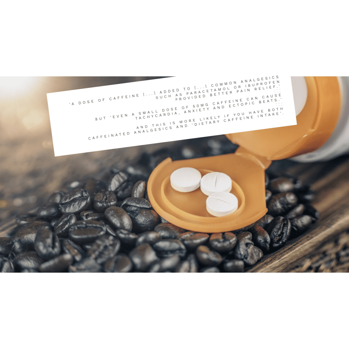
Many of our patients have high temperatures, especially during COVID outbreaks. Some of them are taking over the counter pain relief as required. This is of course recommended, however many of our patients are also tachycardic due at least in part to their high temperatures. When they complain of palpitations, do they need to be aware of the effects of caffeine-paracetamol combinations? Should they be taking this combination at all, or should they simply ease off dietary sources of caffeine while taking analgesics? Here are a few quotes on the subject for discussion. Full references are below. For a fast fact summary, have a look at the featured image below:
A dose of caffeine equivalent to a mug of coffee added to a standard dose of common analgesics such as paracetamol or ibuprofen provided better pain relief. Analgesic plus caffeine increased the number of people who had a good level of pain relief by 5% to 10% compared with analgesic alone
Derry et al, 2014.
But…
When the recommended paracetamol-caffeine dosing regimen is combined with dietary caffeine intake, the resulting higher dose of caffeine may increase the potential for caffeine-related adverse effects such as insomnia, restlessness, anxiety, irritability, headaches, gastrointestinal disturbances and palpitations.
EMC, 2017
Even a small dose of 50 mg caffeine can cause tachycardia, anxiety and ectopic beats. Toxicity is normally seen at doses > 500 mg, but this depends on tolerance.
Take into account dietary and other medicinal sources of caffeine: people may not be aware from the brand name that a particular preparation contains caffeine. Consider whether paracetamol with caffeine is necessary: it cannot be assumed that it will be tolerated in the same way as paracetamol alone.
Reference: National Institute for Health and Care Excellence. (2014). Head injury: assessment and early management (NICE Clinical Guideline 176). Retrieved from https://www.nice.org.uk/guidance/cg176
This has not yet been peer reviewed. Comment and review below 🙂
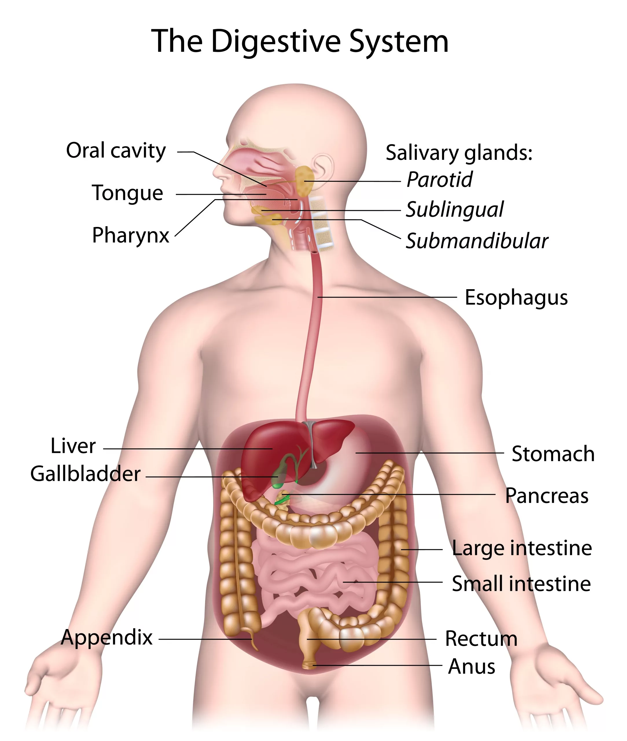
‘The human digestive system consists of the alimentary canal [the gut] and its associated glands, the salivary glands, the liver and the pancreas. The alimentary canal begins at the mouth and ends at the anus. Between the two openings is a long convoluted tube organised into several distinct regions.’
(Boyle and Senior, 2008: 131).
The Short Answer
The digestive system is:
the ‘alimentary canal and its associated glands, the salivary glands, the liver and the pancreas.’ (Boyle and Senior, 2008: 131)
The Extended Answer
Within the alimentary canal are:
The mouth, including the tongue and teeth.
The oesophagus, which ‘carries food from the mouth to the stomach’
The stomach, ‘a muscular bag or sac that stores food’
The small intestine, which is where most digestion and absorption occurs. It includes the:
duodenum
ileum
The large intestine, including the:
appendix
colon, ‘whose main function is to absorb water’
rectum
The anus
(Boyle and Senior, 2008: 130-131)
The digestive system is therefore consisted of all of the above as well as the liver ‘and its adjuncts – the gallbladder and bile ducts’ (Keeton et al, 2020), pancreas, and the salivary glands.
Of course, each component has its own functions and parts. As such, this answer could be extended even further.
References
Boyle, M. And Senior, K. 2008. Human Biology, Third Edition, Collins: London
Collison, P. et al, 2001. Nelson Modular Science 1, Nelson Thornes: Cheltenham
Keeton, W.T. et al, 2020. Human Digestive System, Available Online: https://www.britannica.com/science/human-digestive-system (Accessed 13/09/20)
Digestive system with salivary glands (licensed Adobe image)