If you, like me, have never internalised the whole HISS thing for areas of the heart and are fed up with people telling you it’s easy, or to visualise it, STOP trying. Keep it simple. People think this is simple but it is only for them. Instead, think about where you apply the ECG leads. Visualise that.
V1 and V2 are placed on the sternum. You can think sternum for septal, or ‘high anterior’ (as they are high up on the front of the chest).
Leads v3 and v4 are placed on the front of the chest. They are anterior.
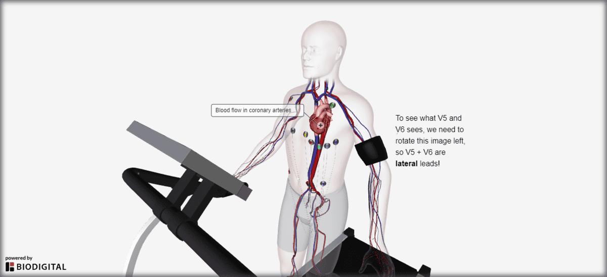
To see where leads V5 and V6 go, you have to turn an image of the heart to the left. They are lateral leads!
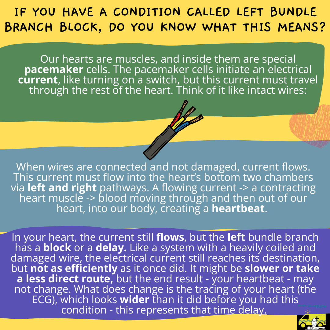
Because of the risk of this, if this is new we are likely to advise you to attend hospital. We may advise you to attend a specialist cardiac hospital, depending on your symptoms and other signs of illness that you might have called us for. If you already know you have the condition, it is worth noting it down and keeping a copy of your ECG trace for any future queries.
If you are a healthcare professional and you are reading this, please let us know what you think by commenting or getting in touch. Would you use this to explain the condition to your patients?
This video demonstrates how to insert an OP airway adjunct to an adult patient. A full video, showing how to size & insert the adjunct, along with information about relevant anatomy, is available on our YouTube channel. For more videos like this, stay tuned to www.article999.co.uk as well as the YouTube, Facebook and Twitter pages.
This is the shorter version of the OPA video. For the long version, featuring the when, where and how of OP airway adjuncts, please click here.
All of the content published by Article 999 follows reputable guidelines and are referenced. This video has not been endorsed by any organisation, author or ambulance trust. You must read the full disclaimer available at www.article999.co.uk/about/ (disclaimer tab) before putting into place anything you read or watch here. Please also remember to check your local guidelines before practicing any of these skills.
Transcript
Remember hand hygiene, bare below the elbows, and gloves in real life! (AACE, 2016)
Having opened the airway using manual airway manouvres and checking it is clear of obstruction, and having already selected the appropriate airway adjunct and size, now insert the adjunct back to front (Pilbery & Lethbridge, 2016) with the bendy part, named ‘the body’ (Beattie, 2005), curved towards the patient’s upper lip and nose. Once the adjunct has reached the soft palate, rotate it 180 degrees and advance it (Pilbery & Lethbridge, 2016). It should now rest in the pharynx (Pilbery & Lethbridge, 2016).
Article 999’s own or licenses purchased via Adobe Stock & Dreamstime
Final note.
Why is it so important to size the adjunct? Because ‘If the airway [device] is too long it may occlude the airway by […] displacing the epiglottis; if too short it will not separate the soft palate or tongue from the posterior wall of the pharnyx’
Gregory, P. & Mursell, I. 2010. ‘Airway management’ in Manual of Clinical Paramedic Procedures, Sussex: Blackwell Publishing, pp. 2-34
This post has been peer reviewed by at least one other individual. Let us know what you think in the comments below.
This video demonstrates how to size and insert an OP airway adjunct and points out relevant airway anatomy. A summary, showing OP airway insertion only, is available on our YouTube channel. For more videos like this, stay tuned to www.article999.co.uk as well as the YouTube, Facebook and Twitter pages.
All of the content published by Article 999 follows reputable guidelines and are referenced. This video has not been endorsed by any organisation, author or ambulance trust. You must read the full disclaimer available at www.article999.co.uk/about/ (disclaimer tab) before putting into place anything you read or watch here. Please also remember to check your local guidelines before practicing any of these skills.
Transcript
Remember hand hygiene, bare below the elbows, and gloves in real life! (AACE, 2016)
Airway adjuncts ‘prevent the tongue from partially or completely obstructing the airway’ (Anaesthesia UK, 2010).
The key parts of the upper airway for this video concern the nasal cavity, the oral cavity, the hard palate, the soft palate (at the back of the mouth), the oropharnyx and the pharynx, which is a ‘muscular membranous channel’ (QA International, 2017) that ‘begins at the base of the skull’ and ‘connects the nasal cavities to the […] oesophagus’ (Pazhaniappan, 2017). It is made up of the nasopharynx, oropharynx, and laryngopharynx (Pazhaniappan, 2017). It is also the location a correctly sized OP airway should sit in.
Use an OP airway on ‘an unresponsive patient’ who does not have a gag reflex (Pilbery & Lethbridge, 2016).
Having opened the airway using manual airway manouvres and checking it is clear of obstruction, now size the adjunct (Pilbery & Lethbridge, 2016). OP airways range from size 000 to 5 (AACE, 2016). To find the right size, measure ‘the vertical distance between the patient’s incisors and the angle of the jaw’ (Pilbery & Lethbridge, 2016), as shown. The flange (Beattie, 2005) should align with the lips and ‘the tip to the angle of the jaw’ (Anaesthesia UK, 2017). When correctly fitted, the OP airway should be just big enough to have the flange (Beattie, 2005) resting over the patient’s lips. Now insert the adjunct back to front (Pilbery & Lethbridge, 2016) with the bendy part, named ‘the body’ (Beattie, 2005), curved towards the patient’s upper lip and nose. Once the adjunct has reached the soft palate, rotate it 180 degrees and advance it (Pilbery & Lethbridge, 2016). It should now rest in the pharynx (Pilbery & Lethbridge, 2016).
Association of Ambulance Chief Executives (AACE). 2016. UK Ambulance Services Clinical Practice Guidelines 2016, Bridgwater: Class Professional Publishing
Pilbery, R. and Lethbridge, K. 2016. Ambulance Care Practice, Bridgwater: Class Professional Publishing QA International, 2017. Respiratory System, Available Online: http://visual.merriam-webster.com/hum… (Accessed 22/08/17)
Article 999’s own or licenses purchased via Adobe Stock & Dreamstime
Final note.
Why is it so important to size the adjunct? Because ‘If the airway [device] is too long it may occlude the airway by […] displacing the epiglottis; if too short it will not separate the soft palate or tongue from the posterior wall of the pharnyx’
Gregory, P. & Mursell, I. 2010. ‘Airway management’ in Manual of Clinical Paramedic Procedures, Sussex: Blackwell Publishing, pp. 2-34
This video has been peer reviewed by one or more people. Let us know what you think by commenting below.
This video shows only the when and how of I-Gels. To view a longer video, which contains the why, what, when and how, click here.
This version has been created to enable those of you who want to refresh yourself on only the need to knows – the use of the i-gel – to do so quickly and easily.
This video has not been endorsed by any organisation, author or ambulance trust. You must refer to local guidelines and read Article 999’s full disclaimer, available at www.article999.co.uk/about/ (disclaimer tab), before putting into place anything you see or read here.
This video has been peer reviewed by 2 or more individuals. No drastic changes have been suggested, so improvements will be made to future posts.