Tag: Airway
-

Short Skills Video – What Happens When We Squeeze the BVM too Hard and Fast?
Full reference available at Article 999’s reference library here: https://airtable.com/appaQWBYHEs4Y6RjF/tblatVkrBMGBMdpRM/viwMSEJkOai9GwYS4/recFg1GuvEjKQZN9f?blocks=hide
-

How do I get the ventilator off the racks?
If you’ve ever felt the frustration of being a student on Day 1 and having to retrieve the ventilator for your crewmate, you might enjoy this GIPHY.
-
What is the relief pressure dial on the ventilator?
This article refers to the workings of Smith’s Medical Pneupac ParaPac and the newer ParaPac Plus. Other transport ventilators may vary in their settings and processes. Please check what unit your ambulance trust has before following the information here.
[tabby title=”Detail”]
The pressure of air inside our lungs is normally just right to not only ensure that negative pressure breathing occurs but also to prevent a shunt (where blood bypasses the lungs) (Hess & Kackmarek, 2014: 431), the oesophageal spinchter from opening, causing air to enter the stomach (Baker, 2012: 1), and a host of other potential problems. Any means of supported ventilation where the patient is not breathing for themselves produces positive pressure ventilation. Not only does this change the way air enters the patient’s lungs while they are ventilated, but it also comes with all of the above risks if any of the settings are incorrect, and more.
Many of the effects ‘are related to mean airway pressure [which is] […] the average pressure applied to the airway during the ventilatory cycle’ (Hess & Kackmarek, 2014: 420) With too ⇑ pressure comes ⇓ venous return (Hess & Kackmarek, 2014: 420) and ⇑ chance of barotrauma (Baker, 2016: 75).
To summarise, the potential negative effects are:
- Blood bypassing the lungs (a shunt) (Hess & Kackmarek, 2014: 431)
- Air entering the stomach (Baker, 2012: 1)
- Venous return decreasing (Hess & Kackmarek, 2014: 420)
- Barotrauma (Baker, 2016: 75)
Because modern ventilators are volume-controlled, preventing a different problem that occurs during pressure-controlled ventilation, there is a set tidal volume which the ventilator will always provide to the patient. To provide this volume, the pressure will constantly increase until the volume is reached (Baker, 2016: 132).
The Means of Avoiding These Problems
In ventilators, the pressure of an oxygen cylinder ‘is lowered through a reducing valve down to 30-90psi’ from 2000psi. It’s then delivered to the patient ‘to a maximum of 40cmh20 or 60cmh20’, (Baker, 2012: 18)*, depending on whether you’re in the UK or the US.
*cmh20 = centimetres of water pressure (SensorsONE, 2017)
Since we can’t have too much pressure there needs to be a way of stopping the ventilator if things get awry. The relief pressure is what this refers to. Via a safety valve (Baker, 2012), otherwise known as ‘exhaust valves’ the supply is cut and excess peak inspiratory pressure (PIP) is released into the atmosphere (Zaconeta et al, 2010), ‘prevent[ing] over-ventilation of the lungs’ (Baker, 2016: 145)
Why would the pressure get this high anyway?
- ‘The patient circuit […] [could be] blocked or kinked’ (Baker, 2016: 142). Best to check it.
- ‘If the lung compliance[*] is low and the airway resistance is high the pressure can rise to high levels’ because, as above, the pressure will keep rising until the set tidal volume is delivered (Baker, 2016: 142).
- The wrong relief pressure settings.
*Compliance ‘refers to the ease of expansion of the lungs and the walls of the thorax’ (Baker, 2012: 8). Ordinarily, according to Baker (2012: 8), the value is 100 cmh20 but when ventilated it’s 40-50cmh20. Physical injuries and medical conditions may further alter this figure.
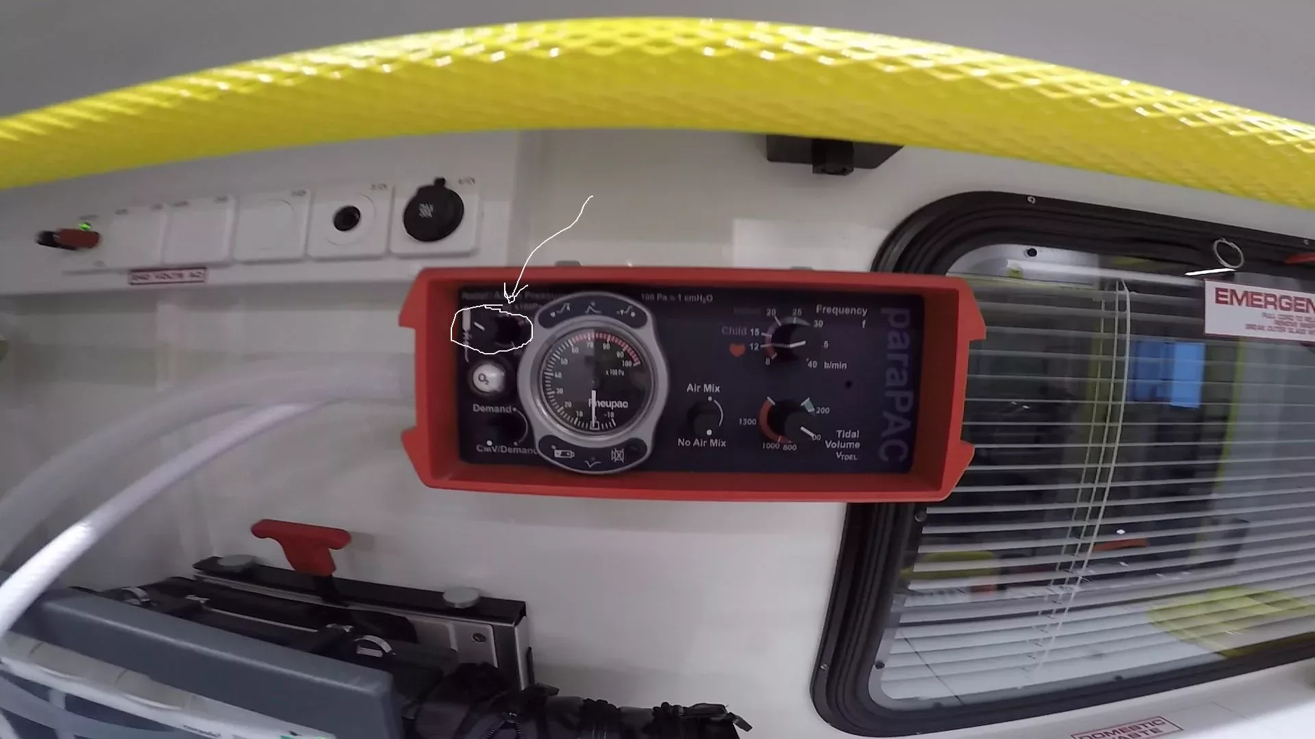
Which dial are you talking about?
This one

The Pneupac ParaPac (Smiths Medical). Photo highlights the Relief Pressure dial. What should I set it to?
40cmh20 if you’re in the UK (Baker, 2012)
Why is the relief pressure set at 40cmh20 in the UK? Why is it 60cmh20 in the US?
Article 999 is struggling to find the answers to these questions. If you know the answers, please get it touch. It would be great to add the information to this resource.

This post is yet to be peer reviewed. Please get in touch if you have any comments. [tabby title=”Super Summary”]
Article 999 previously posted an article on positive pressure ventilation, including its risks. The aim of positive pressure ventilation is to ‘maximise the potential benefit of mechanical ventilation while minimizing the potential for harm’ (Hess & Kackmarek, 2014: 407). The relief pressure dial on the ventilator ensures that the pressure of ventilation is not so high as to cause blood bypassing the lungs (Hess & Kackmarek, 2014: 407), the oesophageal spinchter opening (Baker, 2012: 1), barotrauma (Baker, 2016: 75; Hess & Kackmarek, 2014) and more. The ventilator’s other settings ensure that the patient actually receives ventilation, but the relief pressure setting ensures that a valve drains excess air when settings, poor setup, or the patient’s lung compliance (Baker, 2016: 43) have caused the pressure to get to high.
What should I set it to?
40cmh20 (Baker, 2012)
What should I look out for if the pressure is too high?
Kinked tubing (Baker, 2016: 142) & your settings
Read more in the Detail tab.
[tabby title=”Prefer to see a diagram? Check out the mind-map summary”]

[tabby title=”References”]
References
Baker, D. 2012. Emergency and Transport Ventilation, an introductory guide, Smiths Medical International Limited: Luton
Baker, D.J. 2016. Artificial Ventilation: A Basic Clinical Guide. Springer International: Switzerland
Hess, D.R. & Kacmarek, R.M. 2014. Essentials of Mechanical Ventilation. McGraw Hill Education: New York
SensorsONE, 2017. cmh20 – Centimetres of Water Column at 4 deg C Pressure Unit, Available Online: https://www.sensorsone.com/cmh2o-centimetres-water-column-4-deg-c-pressure-unit/ (Accessed 16/11/17)
Zaconeta, C.A.M. et al, 2010. ‘Evaluation of peak inspiratory pressure and respiratory rate during ventilation of a preterm infant lung model with a self-inflating bag by paramedics of the Fire Department’, Revista Paulista de Pediatria, vol. 28. No. 1.
Not endorsed by but with thanks to Smiths Medical for providing information.
[tabbyending]
-

How to size and insert an OP airway adjunct (insertion only)
This video demonstrates how to insert an OP airway adjunct to an adult patient. A full video, showing how to size & insert the adjunct, along with information about relevant anatomy, is available on our YouTube channel. For more videos like this, stay tuned to www.article999.co.uk as well as the YouTube, Facebook and Twitter pages.
This is the shorter version of the OPA video. For the long version, featuring the when, where and how of OP airway adjuncts, please click here.
Menu
0:08 disclaimer
0:25 Seen this video before? Skip to the main content
0:33 Sizing the OPA
0:38 Inserting the OPA
1:01 References
Disclaimer
All of the content published by Article 999 follows reputable guidelines and are referenced. This video has not been endorsed by any organisation, author or ambulance trust. You must read the full disclaimer available at www.article999.co.uk/about/ (disclaimer tab) before putting into place anything you read or watch here. Please also remember to check your local guidelines before practicing any of these skills.
Transcript
Remember hand hygiene, bare below the elbows, and gloves in real life! (AACE, 2016)
Having opened the airway using manual airway manouvres and checking it is clear of obstruction, and having already selected the appropriate airway adjunct and size, now insert the adjunct back to front (Pilbery & Lethbridge, 2016) with the bendy part, named ‘the body’ (Beattie, 2005), curved towards the patient’s upper lip and nose. Once the adjunct has reached the soft palate, rotate it 180 degrees and advance it (Pilbery & Lethbridge, 2016). It should now rest in the pharynx (Pilbery & Lethbridge, 2016).
Made by www.article999.co.uk
Full disclaimer available at website.
References
Beattie, S. 2005. Placing an oropharnygeal airway, Available Online: http://www.modernmedicine.com/modern-… (Accessed 18/05/17)
Pilbery, R. and Lethbridge, K. 2016. Ambulance Care Practice, Bridgwater: Class Professional Publishing
Music:
Dobroide, 2010. 20091229.ambulance.siren.wav. Available Online: https://freesound.org/people/dobroide… (Accessed 21/08/17)
Productiontrax.com
Images:
Article 999’s own or licenses purchased via Adobe Stock & Dreamstime
Final note.
Why is it so important to size the adjunct? Because ‘If the airway [device] is too long it may occlude the airway by […] displacing the epiglottis; if too short it will not separate the soft palate or tongue from the posterior wall of the pharnyx’
Gregory, P. & Mursell, I. 2010. ‘Airway management’ in Manual of Clinical Paramedic Procedures, Sussex: Blackwell Publishing, pp. 2-34

This post has been peer reviewed by at least one other individual. Let us know what you think in the comments below. -
How to size and insert and OP airway adjunct: Where, When and How?
This video demonstrates how to size and insert an OP airway adjunct and points out relevant airway anatomy. A summary, showing OP airway insertion only, is available on our YouTube channel. For more videos like this, stay tuned to www.article999.co.uk as well as the YouTube, Facebook and Twitter pages.
Menu
0:07 disclaimer
0:24 anatomy
1:04 When?
1:14 How? Sizing
1:54 Insertion
2:26 References
Disclaimer
All of the content published by Article 999 follows reputable guidelines and are referenced. This video has not been endorsed by any organisation, author or ambulance trust. You must read the full disclaimer available at www.article999.co.uk/about/ (disclaimer tab) before putting into place anything you read or watch here. Please also remember to check your local guidelines before practicing any of these skills.
Transcript
Remember hand hygiene, bare below the elbows, and gloves in real life! (AACE, 2016)
Airway adjuncts ‘prevent the tongue from partially or completely obstructing the airway’ (Anaesthesia UK, 2010).
The key parts of the upper airway for this video concern the nasal cavity, the oral cavity, the hard palate, the soft palate (at the back of the mouth), the oropharnyx and the pharynx, which is a ‘muscular membranous channel’ (QA International, 2017) that ‘begins at the base of the skull’ and ‘connects the nasal cavities to the […] oesophagus’ (Pazhaniappan, 2017). It is made up of the nasopharynx, oropharynx, and laryngopharynx (Pazhaniappan, 2017). It is also the location a correctly sized OP airway should sit in.
Use an OP airway on ‘an unresponsive patient’ who does not have a gag reflex (Pilbery & Lethbridge, 2016).
Having opened the airway using manual airway manouvres and checking it is clear of obstruction, now size the adjunct (Pilbery & Lethbridge, 2016). OP airways range from size 000 to 5 (AACE, 2016). To find the right size, measure ‘the vertical distance between the patient’s incisors and the angle of the jaw’ (Pilbery & Lethbridge, 2016), as shown. The flange (Beattie, 2005) should align with the lips and ‘the tip to the angle of the jaw’ (Anaesthesia UK, 2017). When correctly fitted, the OP airway should be just big enough to have the flange (Beattie, 2005) resting over the patient’s lips. Now insert the adjunct back to front (Pilbery & Lethbridge, 2016) with the bendy part, named ‘the body’ (Beattie, 2005), curved towards the patient’s upper lip and nose. Once the adjunct has reached the soft palate, rotate it 180 degrees and advance it (Pilbery & Lethbridge, 2016). It should now rest in the pharynx (Pilbery & Lethbridge, 2016).
Made by www.article999.co.uk
Full disclaimer available at website.
References
Anaesthesia UK, 2010. Guedal Airway, Available Online: http://www.frca.co.uk/article.aspx?ar… (Accessed 22/08/2017)
Association of Ambulance Chief Executives (AACE). 2016. UK Ambulance Services Clinical Practice Guidelines 2016, Bridgwater: Class Professional Publishing
Beattie, S. 2005. Placing an oropharnygeal airway, Available Online: http://www.modernmedicine.com/modern-… (Accessed 18/05/17)
Pazhaniappan, N. 2017. The Pharynx, Available Online: http://teachmeanatomy.info/neck/visce… (Accessed 22/08/17)
Pilbery, R. and Lethbridge, K. 2016. Ambulance Care Practice, Bridgwater: Class Professional Publishing QA International, 2017. Respiratory System, Available Online: http://visual.merriam-webster.com/hum… (Accessed 22/08/17)
Music:
Dobroide, 2010. 20091229.ambulance.siren.wav. Available Online: https://freesound.org/people/dobroide… (Accessed 21/08/17)
Productiontrax.com
Images:
Article 999’s own or licenses purchased via Adobe Stock & Dreamstime
Final note.
Why is it so important to size the adjunct? Because ‘If the airway [device] is too long it may occlude the airway by […] displacing the epiglottis; if too short it will not separate the soft palate or tongue from the posterior wall of the pharnyx’
Gregory, P. & Mursell, I. 2010. ‘Airway management’ in Manual of Clinical Paramedic Procedures, Sussex: Blackwell Publishing, pp. 2-34
This video has been peer reviewed by one or more people. Let us know what you think by commenting below. -
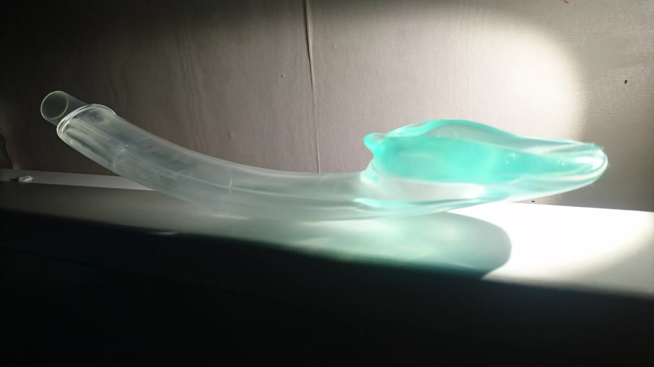
I-Gel: When, How?
This video shows only the when and how of I-Gels. To view a longer video, which contains the why, what, when and how, click here.
This version has been created to enable those of you who want to refresh yourself on only the need to knows – the use of the i-gel – to do so quickly and easily.
This video has not been endorsed by any organisation, author or ambulance trust. You must refer to local guidelines and read Article 999’s full disclaimer, available at www.article999.co.uk/about/ (disclaimer tab), before putting into place anything you see or read here.
This video has been peer reviewed by 2 or more individuals. No drastic changes have been suggested, so improvements will be made to future posts. -
I-Gel: Why, What, When, How?
Over the coming weeks and months, Article 999 aims to build a video library of skills and equipment, and in the future, scenarios. The aims are to make it easier for all operational staff to access refreshers or aid their training, while also enabling students to learn how to find and use equipment.
These videos should add to a resource that provides information that is easy to find and quick to learn from. Importantly, all of our videos have been produced based on publicly available information that is referenced, and all of them have been created by and for operational staff in the UK.
Feedback is welcome. As with all of our content, please remember to check local guidelines and read our full disclaimer before putting into place anything you see or read here.
Presenting one of our videos, I Gel: Why, What, When, How?
This video has been reviewed by one or more individuals. No drastic changes have been suggested but improvements will be made to future videos. Let us know if you have any feedback in the comments below.