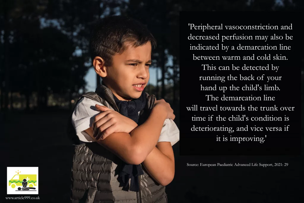
Image above shows a cold child and a quote from the EPALS textbook.
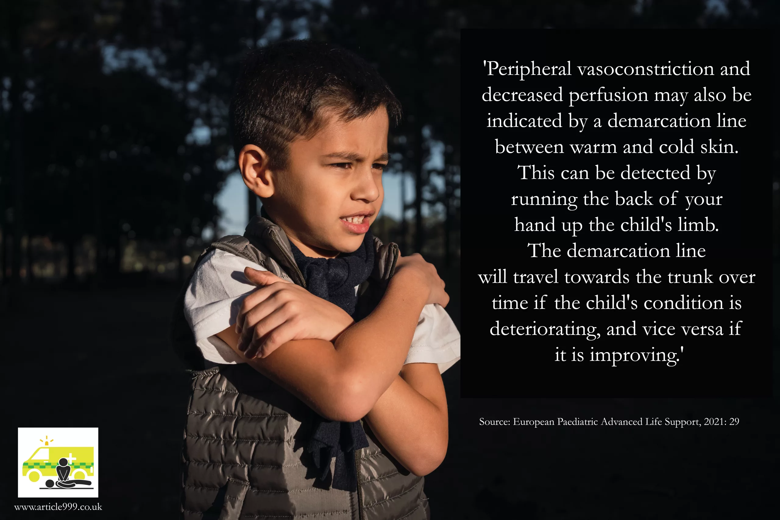
Image above shows a cold child and a quote from the EPALS textbook.
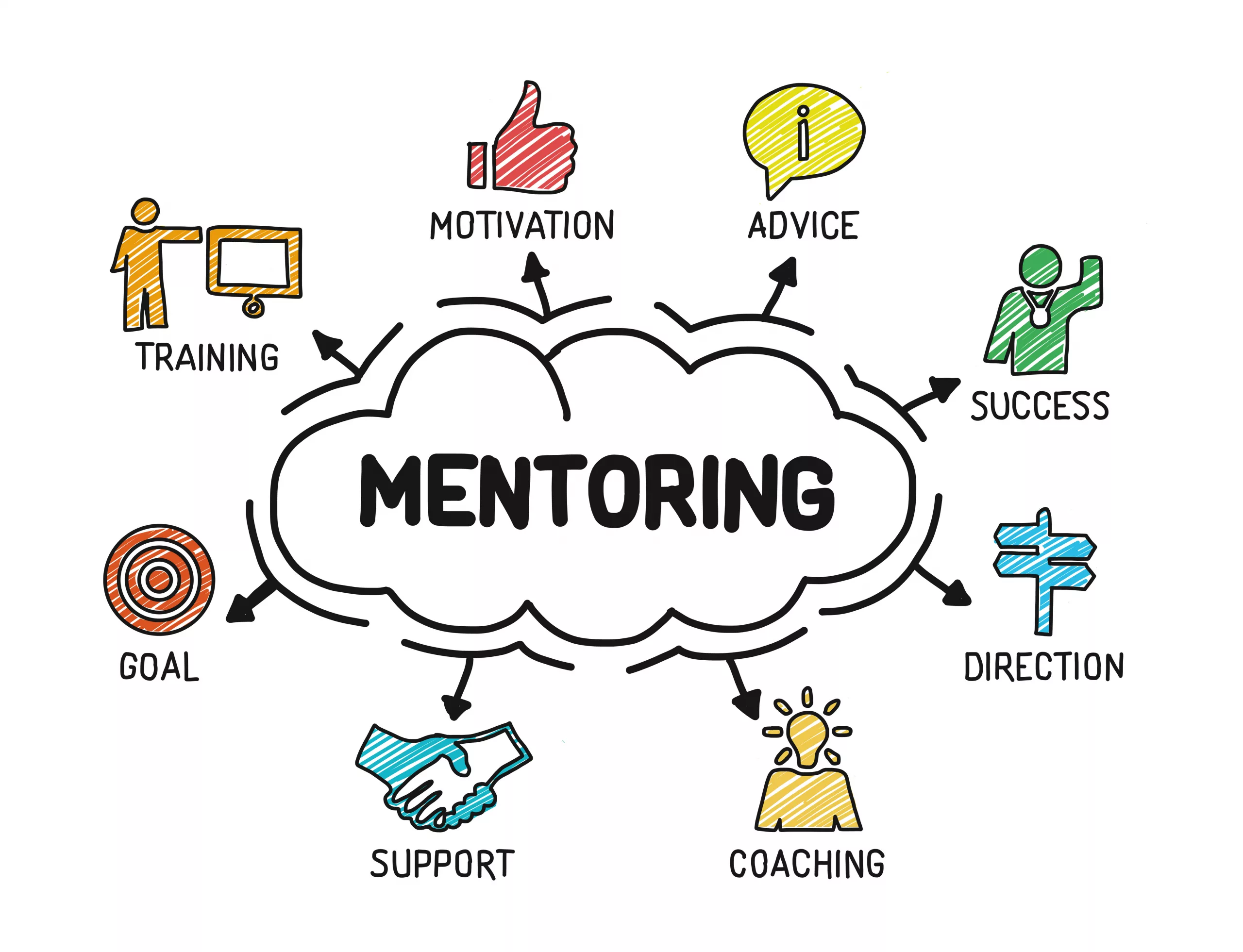
As HCPs we are used to having mentors. We are assigned them in training programmes, trained to become them as part of our progression, and some workplaces offer ‘team leader’ style roles for continued mentorship post-training. But what about when we want advice on accessing career pathways that aren’t immediately available?
What about when we are thinking of reducing hours, changing contracts, trying different Trusts and don’t know anyone currently doing the same?
LinkedIn is a great tool – if people respond, and I have said for some time that if you look for someone doing what you would like to do, you will likely find them. The problem is, that doesn’t mean you can ask them how they got there, what courses were worthwhile (or are now worthwhile amidst constantly moving barriers like Trusts previously wanting level 3 certificates now wanting level 4, and evolving career maps), or how to get anyone in your chosen line of work to answer your enquiries.

Paramedics are now working across a variety of fields, and as I have written recently, I believe that we may soon reach a point like other professions of working a generic few years before choosing specialist paths. However, I am aware that many are trying to work out which path is most suited to them, which one isn’t a dead-end and involves a good mix of a healthy shift pattern, interesting work, and career progression. It isn’t easy to choose when many pathways are new or still being developed. Some appear in one part of the country several months before they pop up in another and as most of us have experienced, the same pathway doesn’t necessarily look or feel the same in every Trust.
I believe that it should be easier for us to find mentors and support each other to answer simple questions like those written above. I hope to soon provide Article 999 mentors who will available to answer questions and offer guidance. These mentors will be working in a variety of specialised roles. They may have specialist interests that have become part of their career. Going forward, they may be able to provide more in-depth career discussions and advice, and some of them may be interested in collaborating on projects and ideas.

We as Paramedics do have the benefit of information and support available from unions and the College of Paramedics, but we don’t have an easy to access resource of helpful, responsive, qualified mentors who are there to help you:

If you are interested in becoming a mentor for Article 999 please email me at article999uk[at]gmail.com with your name, role (and registration number if you are registered), clinical background and experience, area of specialist interest(s), and mentoring experience and qualifications. Please note this is currently voluntary, but committed mentors will be contacted in the future as this project develops and grows. Mentors will also receive a certificate that could be used for CPD portfolios.
If you are interested in being mentored then please let us know by liking this post, commenting and/or sharing it.
“Mentoring is a brain to pick, an ear to listen, and a push in the right direction.”
John C. Crosby
(one of many quotes that describe effective mentoring)

There are so many different recommendations of ECG Interpretation. Some books have 5 steps, some 6, some 9, some 11. Below, I have listed a few common ones in the hope of highlighting the differences – and the similarities. I have tried to focus on rhythm strip analysis only but some methods combine the rhythm strip with the 12 lead analysis. I have only included those that are publicly available, so although some fantastic methods are taught on courses, I can’t reference them and have subsequently omitted them. However, if you have a preference or believe one method should be included below, add it in the comments and it may be featured in a future update.
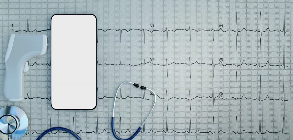
From: Resuscitation Council, Advanced Life Support (2021: 106):
The 6 Steps of rhythm strip analysis:
1. Is there any electrical activity? (Or/and check a pulse)
2. What is the ventricular (QRS) rate?
3. Is the QRS rhythm regular or irregular?
4. Is the QRS complex width normal (‘narrow’) or broad?
—
5. Is atrial activity present?
6. Is atrial activity related to ventricular activity and, if so, how?

From: Charles L. Till, Clinical ECGs in Paramedic Practice (2021: 16):
9 Steps – initially look at lead II, then look at every ECG lead:
ECG Steps
1. What is the rate and rhythm?
2. Are there any P waves and what is their relationship with the QRS complex?
3. What is the duration and morphology of the QRS complex?
4. Is the ST segment isoelectric, depressed or elevated?
5. Are the QT intervals and T waves normal?
Clinical Steps
6. Is the heart generating a palpable pulse of appropriate rate and providing adequate perfusion?
7. Is the rhythm unstable and at risk of deterioration?
8. Does the presenting rhythm support or change your working diagnosis?
9. Are any clinical interventions required?

From: Paul Murray, East of England Ambulance Service, Version 1.0, ECG Recognition – Quick Reference Guide (Feb 2014: 1)
6 steps:
1. ‘QRS Rate’
2. ‘Is the QRS rhythm regular or irregular?’ (and regularly irregular or irregularly irregular?)
3. ‘QRS duration’
4. ‘Are P waves present?’
5. ‘Relationship between P waves and QRS – is there a P wave for each QRS and a QRS for each P wave?’
6. ‘Is the PR interval within normal limits 120-200ms and is it constant?’
(This is basically the same as the RC council method, with one added step; the RC method has one step at the start – a reminder to ensure the leads are connected properly and the patient is alive).
From: Fred Kusumoto & Pam Bernath, ECG Interpretation for Everyone – An On-The-Spot Guide (2012: 33-36):
‘Assess the patient: symptoms and physical examination. Appearance, vital signs, physical examination’ ->
1. Is there a P wave in front of every QRS and is the rate between 50-100 beats per minute?
2. Are the ST segments isoelectric?
Each answer leads you to a different figure – an option to confirm you have a normal ECG, evaluation of ST segments, evaluation of arrhythmias, or confirmation time-critical patients.
The normal ECG should have:
1. Rate should be between 50 and 100bpm
2. A P before every QRS (Positive P in lead II).
3. The QRS in V1 should be narrow an negative i.e. an Rs
4. The ST segment should be isoelectric
5. The T wave should be the same direction as the QRS
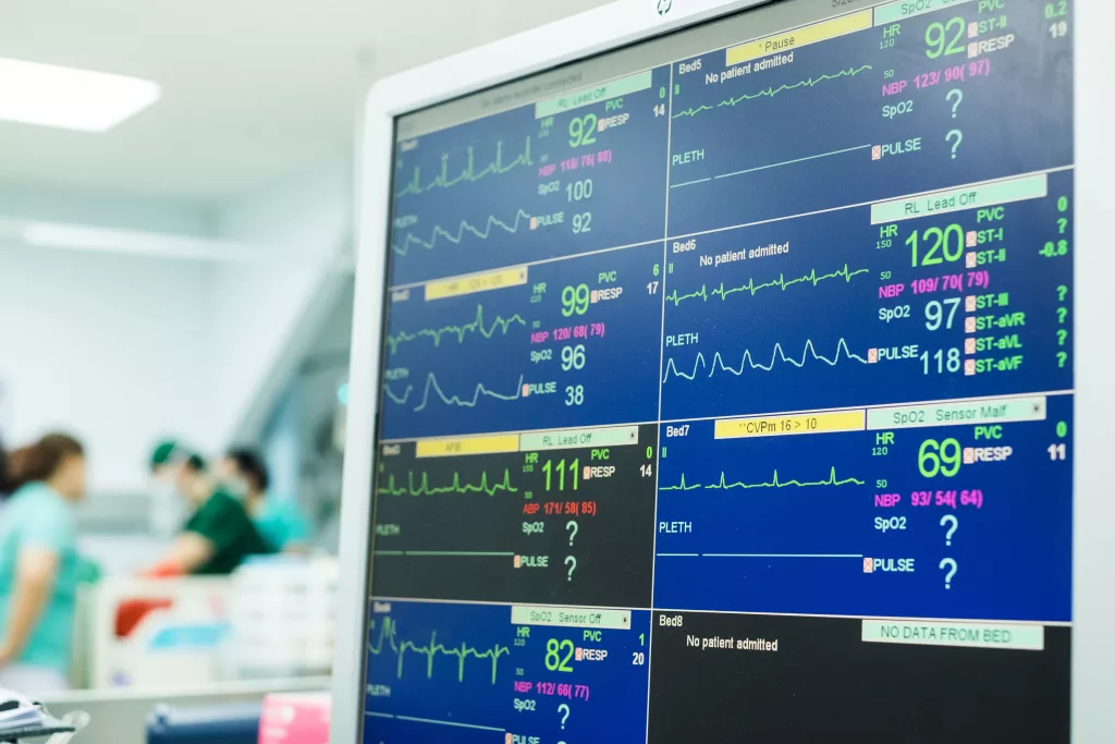
From: Kuhn, Lang, and Wiesbauer, ECG Mastery: The Simplest Way to Learn the ECG (2014: 141-142)
Summarised – 11 steps (taking you from rhythm analysis into 12 lead analysis. Steps included to highlight how some of the steps above are readdressed or appear later altogether here; other points may appear in front):
1. Rhythm. Criteria for sinus rhythm:
Ask: Is it sinus?
2. Heart rate
3. P waves – atrial enlargement present?
4. PR interval
5. QRS axis
6. QRS duration
7. Rotation
8. QRS amplitude
9. QRS infarction signs
10. ST-T segment
11. QT duration, T-U waves
That’s it for today, though you may find other methods at useful courses such as Mark Whitbread’s ECG Bootcamp.
Do you have any recommendations, preferences, or opinions? Add them below!
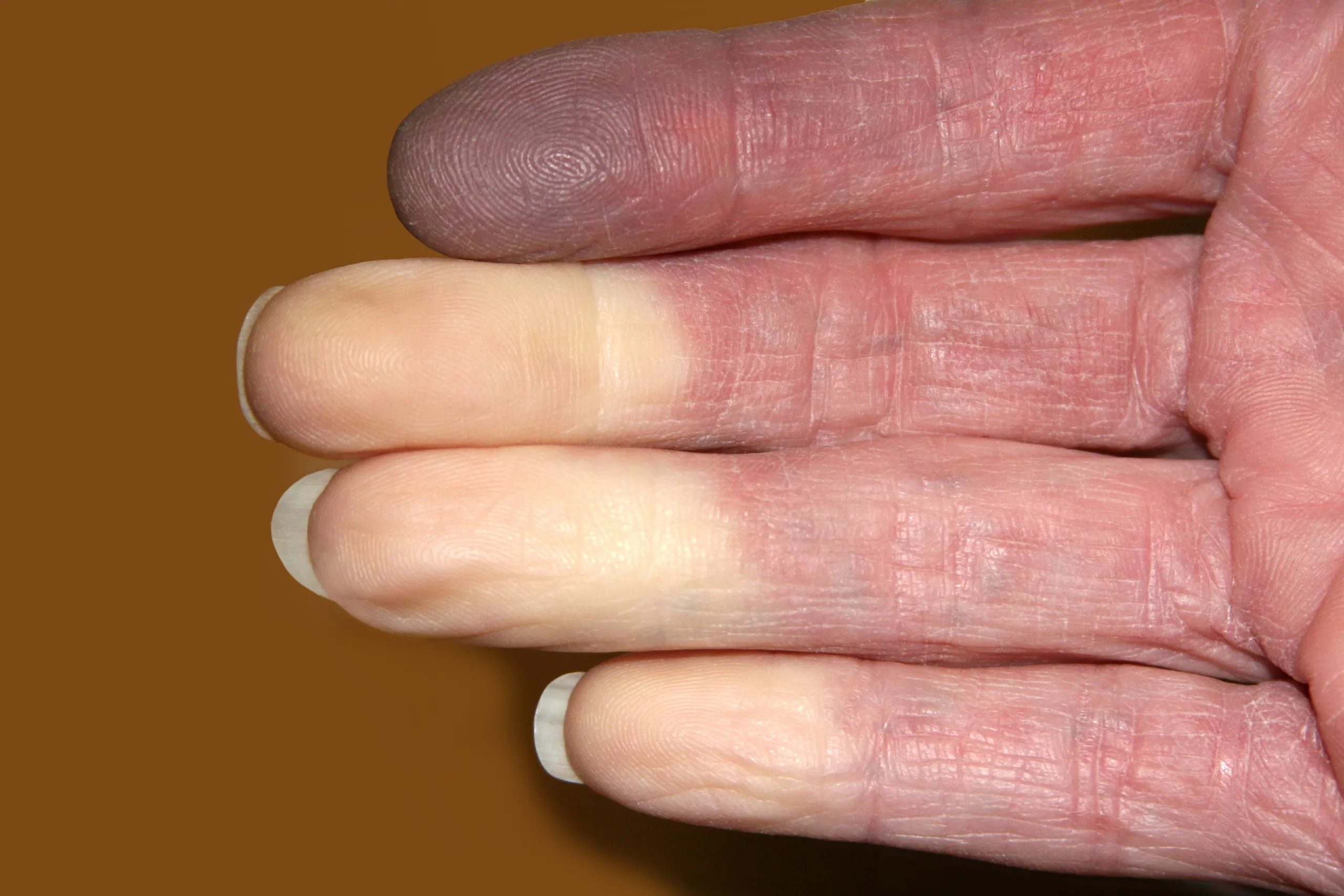
Here’s a list I’ve had for a while but not published: obvious, less obvious, and utterly surprising reasons your alive patient might be pale. References within. Detailed reference list coming soon.

Discoloration of the hands/nails/eyelids:
13. White discoloration of nails – 6 of 155 HIV patients in a 1998 study had this, amongst other more prevalent changes of their nails – https://jamanetwork.com/journals/jamadermatology/fullarticle/189490
14. Anaemia – ‘pallor of the conjunctiva, palm, nail beds or at any site was associated with a significantly lower hemoglobin concentration’ – From <https://academic.oup.com/jn/article/129/9/1675/4721973>
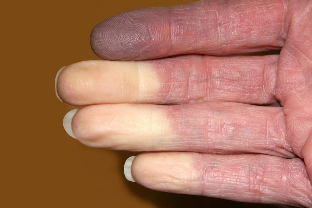
15. Raynaud Phenomenon – leading to white/yellow/purple fingers, temporarily – http://www.cmaj.ca/content/188/15/E396
Red herring
16. Rigor mortis and liver mortis in an alive patient who was suffering from a dissecting abdominal aorta: https://content.sciendo.com/view/journals/sjfs/22/1/article-p11.xml
Have I missed any? Add in the comments below —>
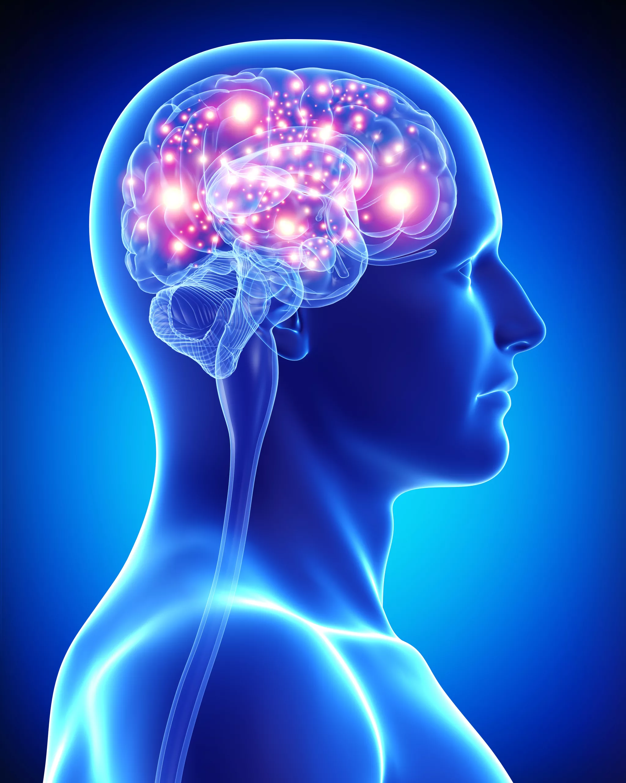
Earlier this year I had the pleasure of speaking with a patient with Functional Neurological Disorder (FND). Ailsa reached out to the College of Paramedics after experiencing an unsettling mixture of treatment by healthcare professionals in a variety of settings. She hopes to encourage healthcare clinicians to learn more about her condition, a functional neurological disorder. Our understanding of this group of conditions is currently being reshaped, so I agree it is another area in which we must stay up-to-date.
The publication of this post on Article 999 comes at a timely moment as just two days ago I read here that a study published in Epilepsy and Behaviour has documented MRI changes in patients with functional seizures. This furthers the point that what science and medicine thought it knew about this – and perhaps other conditions – while not fiction, is also not yet fact.
Members of the College of Paramedics can read the published article in last month’s issue of Paramedic INSIGHT or online here.

| Sign, symptom, or condition | Sign of, and Treatment | Reference |
| Sunken eyes | Dehydration; Patient may need referral to Doctor or urgent rehydration, depending on severity. Patient may benefit from blood tests | Johnson and Smith, 2012: 94. |
| High NEWS2 Score | Sepsis; patient may need antibiotics and emergency treatment | Royal College of Physicians, 2017 |
| Covid-19 | The diarrhoea could be a sign of deterioration, but the evidence for this is weak | Amico et al, 2020 |
| Female and increased age | Increased risk of dehydration | Rowat et al, cited in Sweetser, 2012 |
| Heart or kidney failure; take steroids | Increased risk of overhydration; patient may require emergency treatment | Sweetser, 2012; WHO, 2021. |
| Dizziness | Dehydration; patient may require urgent rehydration | Sweetser, 2012; WHO, 2021 |
| Confusion; seizures | Dehydration or overhydration; electrolyte imbalances; patient may require emergency treatment and urgent rehydration | Sweetser, 2012; WHO, 2021 |
| Type 2 Diabetes; metformin | Metformin can cause the symptoms; diabetes is a risk factor due to potential difficulties managing the condition | Johnson and Smith, 2012: 96 |
| Immunosuppressed | Risk of deterioration | Johnson and Smith, 2012: 96 |
| Bowel disease such as ulcerative colitis or diverticulitis | May indicate more severe illness | Johnson and Smith, 2012: 96 |
| Blood in stools | May be an indication of abdominal bleeding | Johnson and Smith, 2012: 96 |

If you, like me, struggle to understand the action potential, here is a great video from UBC Medicine, which explains the concept with dominoes. Check it out below.

For mentors/PPEDs, PHEM feedback facilitators, ALS instructors, teachers, and those interested in responding better to feedback (and in doing so, giving better feedback), here’s one for you. This is a summary in quotes of the ‘Thanks for the Feedback’ book, picking up on some key points. The full reference is available in our new reference database here: https://airtable.com/shrmbfb43bwcyYtPG
This video took about 2 hours of an evening to produce and was created by Article 999’s founder, Louise, using a great software program purchased out of pocket. The actual reading and saving of quotes has taken [insert actual time] far too long and a similar amount of time will be needed for other books. If you like Article 999’s content, let us know by buying us a coffee here or clicking on the coffee symbol in the bottom right corner.

In 1999, Anna Bagenholm survived a cardiac arrest after a prolonged down-time and a temperature of just 13.7c. The circumstances of her survival continue to make international news and to inform and inspire research about target temperature management. There have been very few documented cases of survival from such extreme hypothermia, especially with minimal neurological impact. Consider the statistics on neurological impact from ordinary cardiac arrests, with less down-time, and Anna’s case becomes even more extraordinary. But does her survival mean we should target hypothermia in cardiac arrest patients? Should we target hypothermia in ROSC? Let’s have a look at the case and the research that has followed:
Situation: 29 Year Old Female (Anna Bagenholm) is submerged head-first in a hole in the ice in a mostly frozen stream for 80 minutes.
Background: She has no medical history. She has slid down an icy slope while skiing with colleagues.
Assessment: She was conscious for the first 40 minutes, but has been unconscious for 40 mins since. After recovery, she is in cardiac arrest. Her ECG shows asystole. During the resuscitation attempt, the following is also learned:
She is warmed and the resuscitation team are watching an echo of her heart, which begins to move. ROSC was not gained until 4 ½ hours after she fell, and she spent 35 days on life support.
Questions: What would your expectations be of her ROSC management? What would your expectations of her survival and neurological impact be?
Recommendation — today:
(Resuscitation Council, 2021)
Decision/Outcome: Not only does Anna survive, her long-term neurological impact is minimal. Have a look at the video:
Have a look at the results of some recent studies into target temperature management, below:
| Year | In or out of hospital study | Presenting rhythm | Targets | Outcome |
| 2002 | OHCA | Shockable | 33 and 37 | Positive for survival and neurological impact |
| 2010 | OHCA | Shockable | Cooled IV fluids (No target temperature listed) | No difference |
| 2013 | OHCA | Both shockable and non-shockable, but presumed cardiac cause | 33 and 36 | No difference |
| 2014 | OHCA | Both shockable and non-shockable | 2L of normal saline at 4 degrees, vs standard care | No difference |
| 2018 | OHCA | Shockable | 32, 33, 34 | No difference |
| 2019 | OHCA and IHCA | Non-shockable | 33 vs 37.5 | Improved survival and good neurological outcome (CPC = 1 or 2) |
| 2021 | OHCA | Presumed cardiac cause | 33 vs 37.5 | No difference |
Why can’t controlled studies replicate the kind of outcome that Anna had? Share your thoughts below, or on our Facebook or Twitter pages.
References (excluding videos and direct links above):
Rasmussen, T. P. and Girotra, S. 2021. A Contemporary Update on Targeted Temperature Management, Available at: https://www.acc.org/latest-in-cardiology/articles/2021/11/09/13/16/a-contemporary-update-on-targeted-temperature-management (Accessed 15/05/2022).
Resuscitation Council, 2021. Post-Resuscitation Care Guidelines, Available at: https://www.resus.org.uk/library/2021-resuscitation-guidelines/post-resuscitation-care-guidelines (Accessed 15/05/2022).