Category: Clinical
-
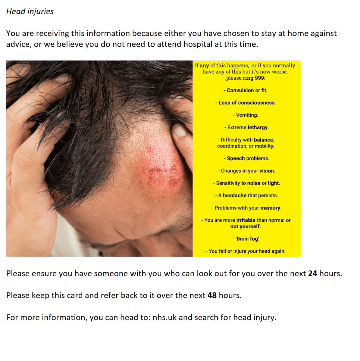
Head Injury: Worsening Advice
The below is an information card designed for patients to receive should ambulance crews need to non-convey. This information is based on the NICE guidelines – https://www.nice.org.uk/guidance/ng232 (2023). This card has not yet been peer reviewed – please add your feedback on the content and presentation below. If you are not a medic, please add your feedback on presentation below.
-

Intercostal Recession in Children
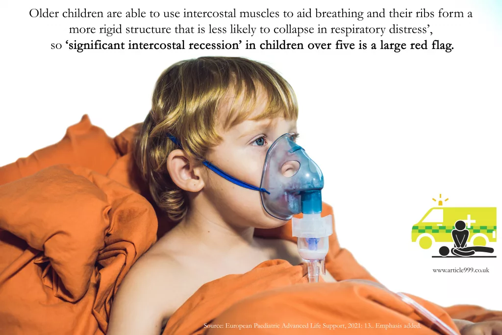
Image with a quote about intercostal recession in children over 5. -
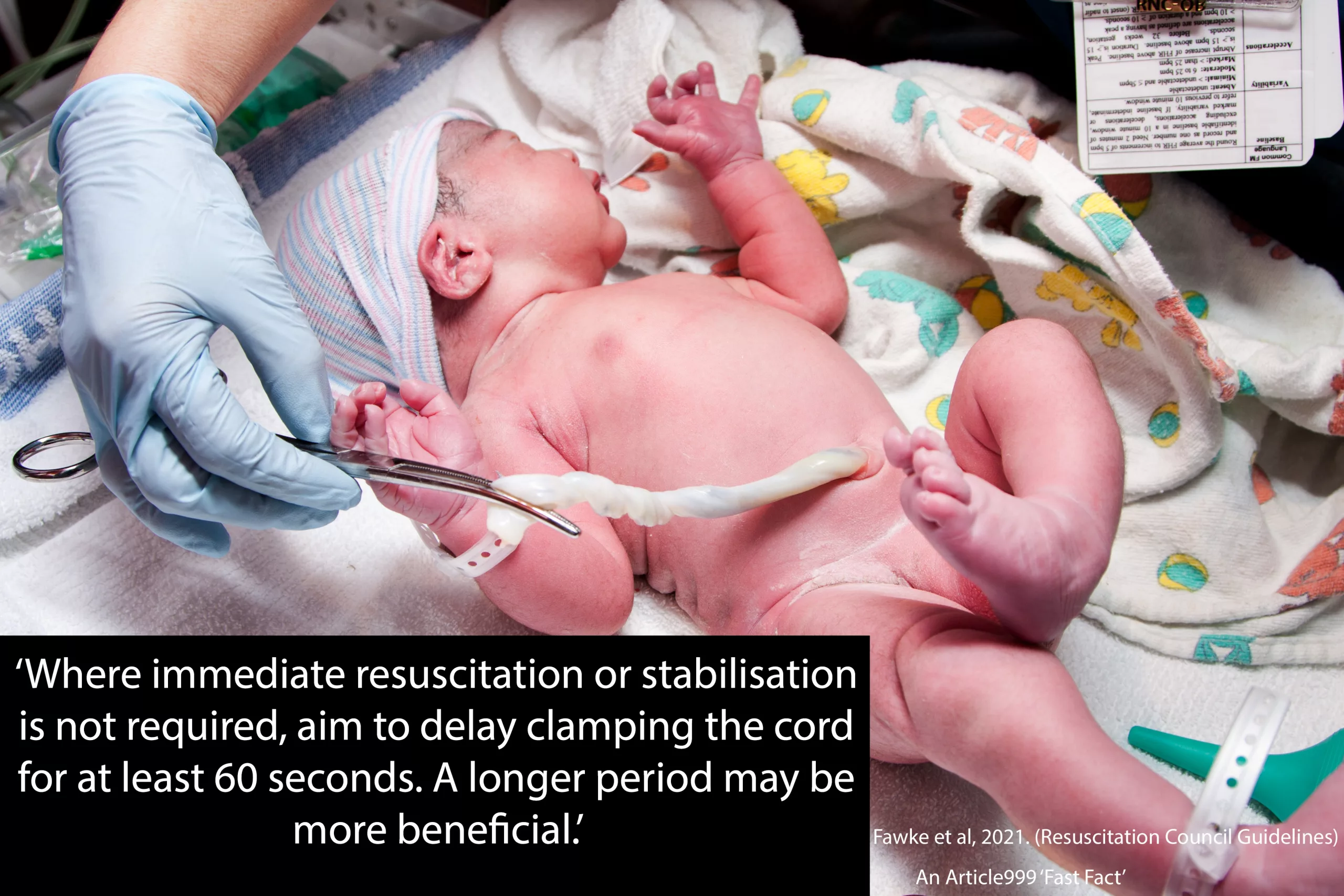
Medics: Should We Delay Cord Clamping? How Long?
Photo containing quote from Resus Council about delayed cord clamping. -
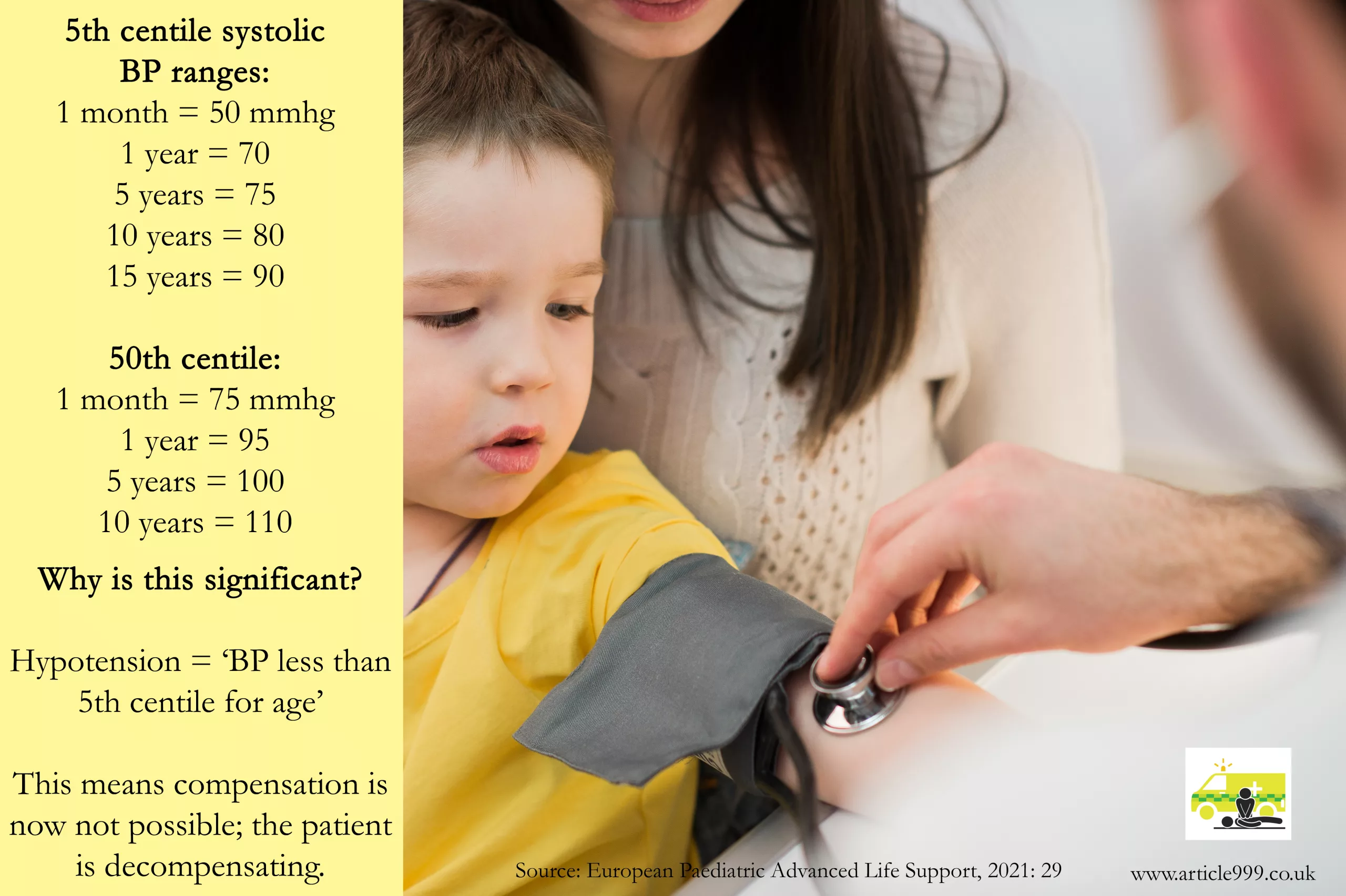
The Decompensating Child: Blood Pressure Ranges to Watch
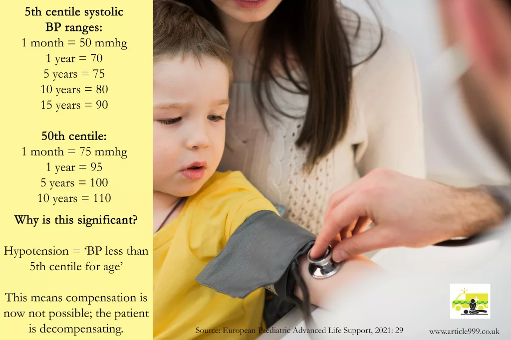
Image showing the 5th and 50th centile blood pressure ranges for children from 1 month-15 years. -

Lacerations – Criteria to Inform Referral Needs in Stable Patients
In your stable patients with lacerations, who do not have any urgent or troublesome A-D problems and who have not sustained injury through any great mechanism, what else should you be looking for in your assessment of their injury? What are the risk factors, according to different guidelines or authors?
While some of these elements may not be enough to refer a patient to secondary care on their own, they should help to increase your risk assessment during safe discharges. In combination, many of these factors should raise flags.
Depth Size Risk Factors Area There is limited evidence regarding the depth of wounds. The emphasis is on the structures that deep wounds would impact. For example, vascular damage, and nerve, tendon, or bony injuries (NICE, 2021). More than 5cm (NICE, 2021). Two or more of:
– Malaise
– Fever
– Rigors
– Contamination of site with body fluids, soil, faeces, or/and pus.
– Patient older than 65 years
– Diabetes
– Jagged wound edges
– Signs of tetanus
(NICE, 2021)Any of:
– Face
– Palm – and there is a potential infection.
– Joint with cellulitis.
(NICE, 2021).Contamination from unknown object (e.g. knife/glass) (NICE, 2021). Presence of necrotic tissue or slough, which could delay healing. Granulation tissue can also delay wound healing, but all can be prevented with appropriate dressings (Wilson, 2012: 11). Wounds older than 24 hours (Newman and Mahdy, 2021). Bites (Newman and Mahdy, 2021). Lacerations: A Collection of Risk Factors and Specific Criteria to Inform Referral Needs in Stable Patients. Have you found more, or would you like to add some from your own experience? Add these in the comments below, and let us know what you think about this post.
References
Newman, R.K. and Mahdy, H. (2021) ‘Laceration’, Treasure Island. Available at: https://www.ncbi.nlm.nih.gov/books/NBK545166/ (Accessed 18/11/2021)
NICE. (2021). Lacerations. Available at: https://cks.nice.org.uk/topics/lacerations/ (Accessed 18/11/2021).
Wilson, M. (2012) ‘Understanding the basics of wound assessment’. Wounds Essentials. 2. Pp. 8-12. Available at: https://www.wounds-uk.com/resources/details/wound-essentials-72-understanding-the-basics-of-wound-assessment (Accessed 28/12/2021).
-

Methods of ECG Interpretation
There are so many different recommendations of ECG Interpretation. Some books have 5 steps, some 6, some 9, some 11. Below, I have listed a few common ones in the hope of highlighting the differences – and the similarities. I have tried to focus on rhythm strip analysis only but some methods combine the rhythm strip with the 12 lead analysis. I have only included those that are publicly available, so although some fantastic methods are taught on courses, I can’t reference them and have subsequently omitted them. However, if you have a preference or believe one method should be included below, add it in the comments and it may be featured in a future update.
Method one:
From: Resuscitation Council, Advanced Life Support (2021: 106):
The 6 Steps of rhythm strip analysis:
1. Is there any electrical activity? (Or/and check a pulse)
2. What is the ventricular (QRS) rate?
3. Is the QRS rhythm regular or irregular?
4. Is the QRS complex width normal (‘narrow’) or broad?
—
5. Is atrial activity present?6. Is atrial activity related to ventricular activity and, if so, how?

Method Two:
From: Charles L. Till, Clinical ECGs in Paramedic Practice (2021: 16):
9 Steps – initially look at lead II, then look at every ECG lead:
ECG Steps
1. What is the rate and rhythm?
2. Are there any P waves and what is their relationship with the QRS complex?
3. What is the duration and morphology of the QRS complex?4. Is the ST segment isoelectric, depressed or elevated?
5. Are the QT intervals and T waves normal?
Clinical Steps
6. Is the heart generating a palpable pulse of appropriate rate and providing adequate perfusion?
7. Is the rhythm unstable and at risk of deterioration?
8. Does the presenting rhythm support or change your working diagnosis?
9. Are any clinical interventions required?
Method Three:
From: Paul Murray, East of England Ambulance Service, Version 1.0, ECG Recognition – Quick Reference Guide (Feb 2014: 1)
6 steps:
1. ‘QRS Rate’
2. ‘Is the QRS rhythm regular or irregular?’ (and regularly irregular or irregularly irregular?)
3. ‘QRS duration’
4. ‘Are P waves present?’
5. ‘Relationship between P waves and QRS – is there a P wave for each QRS and a QRS for each P wave?’
6. ‘Is the PR interval within normal limits 120-200ms and is it constant?’
(This is basically the same as the RC council method, with one added step; the RC method has one step at the start – a reminder to ensure the leads are connected properly and the patient is alive).
Method Four
From: Fred Kusumoto & Pam Bernath, ECG Interpretation for Everyone – An On-The-Spot Guide (2012: 33-36):
‘Assess the patient: symptoms and physical examination. Appearance, vital signs, physical examination’ ->
1. Is there a P wave in front of every QRS and is the rate between 50-100 beats per minute?
2. Are the ST segments isoelectric?
Each answer leads you to a different figure – an option to confirm you have a normal ECG, evaluation of ST segments, evaluation of arrhythmias, or confirmation time-critical patients.
The normal ECG should have:
1. Rate should be between 50 and 100bpm2. A P before every QRS (Positive P in lead II).
3. The QRS in V1 should be narrow an negative i.e. an Rs
4. The ST segment should be isoelectric
5. The T wave should be the same direction as the QRS

Method Five:
From: Kuhn, Lang, and Wiesbauer, ECG Mastery: The Simplest Way to Learn the ECG (2014: 141-142)
Summarised – 11 steps (taking you from rhythm analysis into 12 lead analysis. Steps included to highlight how some of the steps above are readdressed or appear later altogether here; other points may appear in front):
1. Rhythm. Criteria for sinus rhythm:- Are the P waves positive in I and II?
- Is there a QRS complex after each P wave?
- Are the PR intervals constant?
- Are the RR intervals constant?
Ask: Is it sinus?
2. Heart rate
3. P waves – atrial enlargement present?
4. PR interval
5. QRS axis
6. QRS duration
7. Rotation
8. QRS amplitude
9. QRS infarction signs
10. ST-T segment
11. QT duration, T-U waves
That’s it for today, though you may find other methods at useful courses such as Mark Whitbread’s ECG Bootcamp.
Do you have any recommendations, preferences, or opinions? Add them below!
-
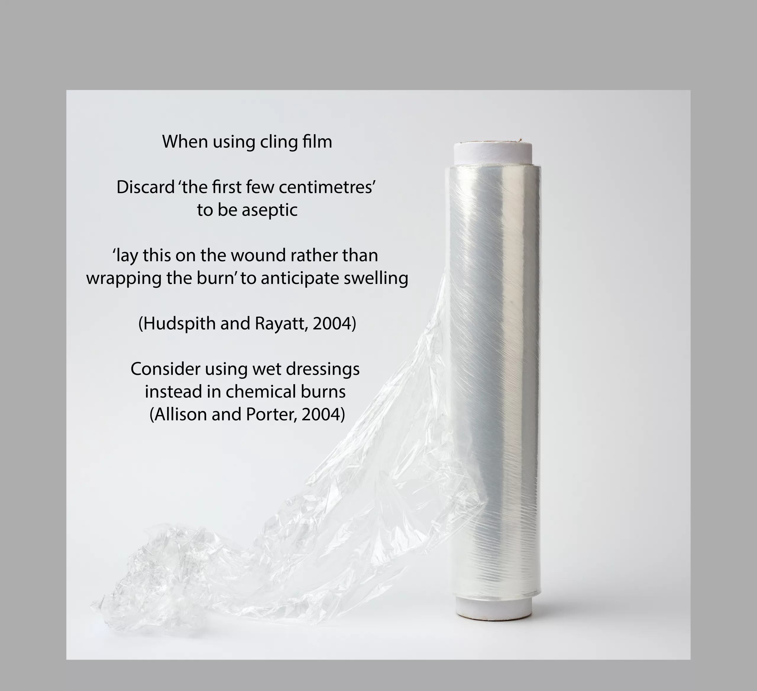
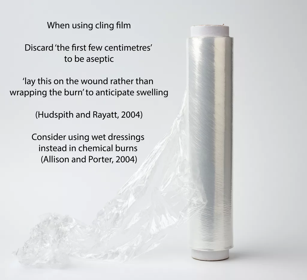
How to Use Cling Film to Treat Burns (Fast Fact)
Fast Fact posts are designed to provide quick and easy ways to revise key topics – view a photo with a quote/fact by simply scrolling through the category. Refresh yourself during those middle-of-the-night breaks when you would have aimlessly scrolled through your phone. There is no need to read a lengthy post to do a little CPD. For more fast facts, head here.