Reference: National Institute for Health and Care Excellence. (2014). Head injury: assessment and early management (NICE Clinical Guideline 176). Retrieved from https://www.nice.org.uk/guidance/cg176
This has not yet been peer reviewed. Comment and review below 🙂
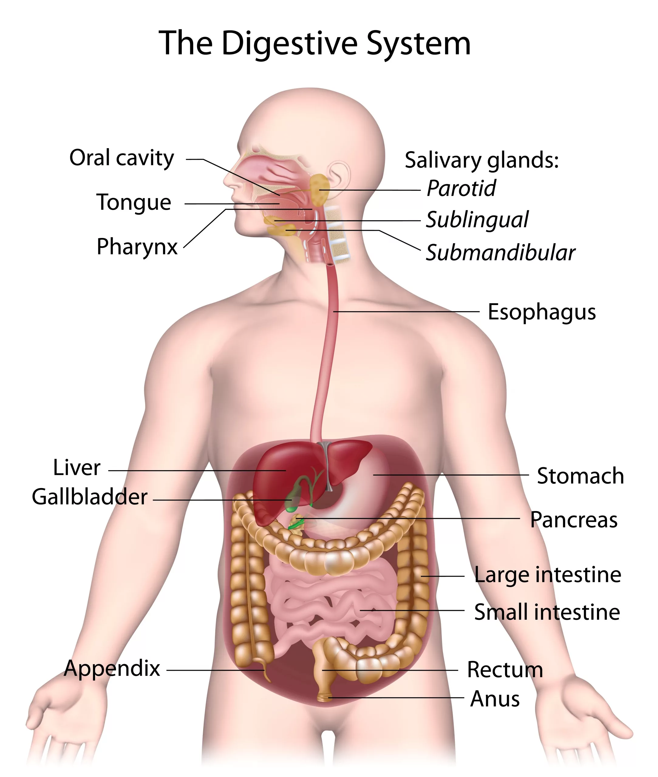
‘The human digestive system consists of the alimentary canal [the gut] and its associated glands, the salivary glands, the liver and the pancreas. The alimentary canal begins at the mouth and ends at the anus. Between the two openings is a long convoluted tube organised into several distinct regions.’
(Boyle and Senior, 2008: 131).
The Short Answer
The digestive system is:
the ‘alimentary canal and its associated glands, the salivary glands, the liver and the pancreas.’ (Boyle and Senior, 2008: 131)
The Extended Answer
Within the alimentary canal are:
The mouth, including the tongue and teeth.
The oesophagus, which ‘carries food from the mouth to the stomach’
The stomach, ‘a muscular bag or sac that stores food’
The small intestine, which is where most digestion and absorption occurs. It includes the:
duodenum
ileum
The large intestine, including the:
appendix
colon, ‘whose main function is to absorb water’
rectum
The anus
(Boyle and Senior, 2008: 130-131)
The digestive system is therefore consisted of all of the above as well as the liver ‘and its adjuncts – the gallbladder and bile ducts’ (Keeton et al, 2020), pancreas, and the salivary glands.
Of course, each component has its own functions and parts. As such, this answer could be extended even further.
References
Boyle, M. And Senior, K. 2008. Human Biology, Third Edition, Collins: London
Collison, P. et al, 2001. Nelson Modular Science 1, Nelson Thornes: Cheltenham
Keeton, W.T. et al, 2020. Human Digestive System, Available Online: https://www.britannica.com/science/human-digestive-system (Accessed 13/09/20)
Digestive system with salivary glands (licensed Adobe image)
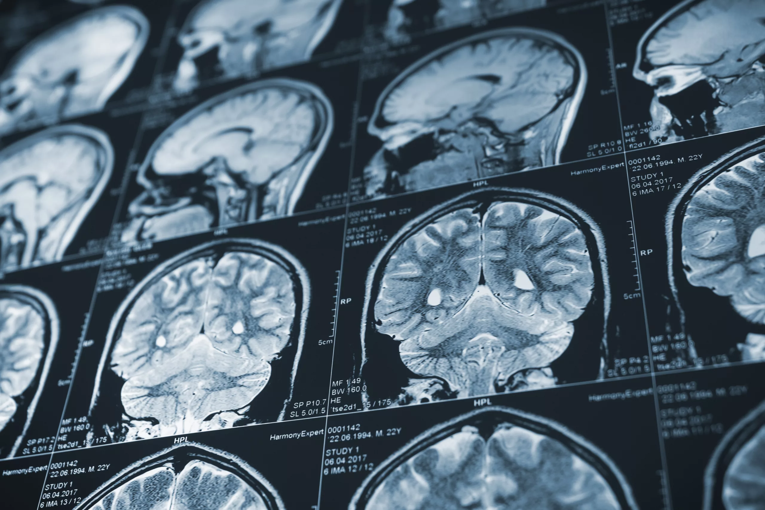
This post is based on the NICE Clinical Guidelines, 2014. One part of their guidelines was in turn updated in 2019.
This post shows the key points and quotes as they relate to frontline ambulance staff; anything not relevant has not been included, but you may read more by following the link to the guidelines here.
Remember to read our disclaimer before putting into practice anything you see, hear, or read here. Also remember to check your local guidelines and the scope of practice for your role before putting any of this into practice.
Article 999: Simplifying the Long Stuff; Presenting the Relevant Stuff; Refreshingyou on the Important Stuff.
www.article999.co.uk
Definition of Head Injuries
‘any trauma to the head other than superficial injuries to the face.’ p6
National Institute for Health and Care Excellence [NICE], 2014: 6
Statistics
‘Head injury is the commonest cause of death and disability in people aged 1-40 years in the UK.’
NICE, 2014: 6
‘The incidence of death from head injury is low, with as few as 0.2% of all patients attending emergency departments with a head injury dying as a result of this injury.’
NICE, 2014: 6
‘the majority of fatal outcomes are in the moderate (GCS 9-12) or severe (GCS 8 or less) head injury groups’
NICE, 2014: 6
When might adults need to attend A&E for a CT head scan following a head injury?
‘For adults who have sustained a head injury and have any of the following risk factors’
Reduced GCS, ‘less than 13’ initially, or ‘less than 15 at 2 hours after the injury on assessment in the emergency department’*
suspected skull fracture of any type
‘post-traumatic seizure’
Neurological deficit
‘More than 1 episode of vomiting’
–> This should happen within one hour of identifying the situation.
(NICE, 2014: 10)
*Consider ‘the pre-injury baseline GCS may be less than 15. Establish this where possible’ (NICE, 2014: 19).
If the patient is on anticoagulants and has none of the above, they should have a CT head ‘within 8 hours of the injury’. (NICE, 2014: 12)
When might adults need to attend A&E for a CTcervical spinefollowing a head injury?
‘The patient has been intubated’
‘The patient is having other body areas scanned for head injury
‘…there is clinical suspicion of cervical spine injury and any of the following apply:
=> 65
‘dangerous mechanism of injury’
neuro deficit
‘paraesthesia in the upper or lower limbs’
(NICE, 2014: 13)
When might children need to attend hospital for a CT head scanfollowing a head injury?
Any of:
‘Suspicion of non-accidental injury’
‘Post-traumatic seizure but no history of epilepsy’
Reduced GCS <14 initially, <15 2 hours later
For under 1 year olds, Reduced GCS <15 on the paediatric scale
Suspected skull fracture of any type
Neuro deficit
For under 1 year olds, ‘presence of bruise, swelling or laceration of more than 5 cm on the head’
(NICE, 2014: 11)
If a child has ‘more than one’ of these, he/she should have a CT scan within an hour:
‘Loss of consciousness lasting more than 5 minutes (witnessed)’
‘Abnormal drowsiness’
Vomiting x3 or more episodes
‘Dangerous mechanism of injury (high-speed road traffic accident either as pedestrian, cyclist or vehicle occupant, fall from a height of greater than 3 metres, high-speed injury from a projectile or other object)’
‘Amnesia […] lasting more than 5 minutes’
(NICE, 2014: 11)
Note: If the child has just one of these, he/she ‘should be observed for a minimum of 4 hours after the head injury’ and if he/she then develops more of the above, a CT is warranted. (NICE, 2014: 12)
The rules regarding CT cervical spine scans are different in children, compared to adults, due to the risk of radiation to their thyroid.
‘Consider or suspect abuse as a contributory factor to or cause of head injury in children’ (NICE, 2014: 7)
For what other reasons should an adult or child with a head injury attend A&E?
loss of consciousness
‘Amnesia for events before or after the injury’
‘Persistent headache since the injury’
‘Any vomiting episodes since the injury’ – but NICE advise considering the causes of single vomiting episodes in those under 12
‘Any seizure since the injury’
‘Any previous brain surgery’
‘A high-energy head injury’
‘Any history of bleeding and clotting disorders’
Anticoagulants
‘Current drug or alcohol intoxication’
Safeguarding issues
‘Continuing concern by the professional about the diagnosis’ (NICE, 2014: 17-18)
And:
Patients who, 48 hours later, have ‘any persistent complaint relating to the initial head injury’ (NICE, 2014: 23)
Also, ‘depending on judgement of severity:’
‘irritability or altered behaviour’
Other ‘Visible trauma to the head […] of concern to the professional’
‘No one is able to observe the injured person at home’
‘Continuing concern by the injured person or their family or carer about the diagnosis’ (NICE, 2014: 18)
What else should I consider in my assessment and treatment of a patient with a head injury?
For adults, NICE recommends ‘managing their care according to clear principles and standard practice’ as in the ATLS and PHTLS courses, and the JRCALC for adults, and the APLS and PHPLS courses for children. There are others referenced, but Article 999 has included the most relevant here. (NICE, 2014: 19-20)
‘Manage pain effectively because it can lead to a rise in intracranial pressure’ (NICE, 2014: 21)
‘Ascribe depressed conscious level to intoxication only after a significant brain injury has been excluded’ (NICE, 2014: 21-22)
Pre-alert patients with a reduced GCS, especially of <8. They will most likely need anesthetist or critical care involvement (NICE, 2014: 21-22)
What’s worth bearing in mind during hospital-neuroscience unit transfers of patients with head injuries?
Patients who have a GCS of less than 8 should be intubated
Before transporting, make sure to stabilise the patient and ensure monitoring is attached
A patient ‘with persistent hypotension’ should not be transported until they are ‘stabilised’ (NICE, 2014: 32-33)
During these transfers, patients ‘should be accompanied by a doctor with appropriate training and experience in the transfer of patients with acute brain injury. […] Patients requiring non-emergency transfer should be accompanied by appropriate clinical staff.’
NICE, 2014: 32-33
Reference
National Institute for Health and Care Excellence. (2014). Head injury: assessment and early management (NICE Clinical Guideline 176). Retrieved from https://www.nice.org.uk/guidance/cg176
In a 2010 study of nursing care in an Italian hospital, researchers found that just ‘40% of nursing activities observed were included in the nursing records’ (Marinis et al, 2010). Although this study was about nurses, not prehospital medics, it indicates the potential for missing key information in our documentation. A variety of models exist to help ensure that all the useful information is in your paperwork by the time it’s finalised & to ensure that it’s clear and concise…
Before the methods are introduced, make sure to note the following:
There is not just one method
Patient report forms vary across Trusts. Essential (required) criteria varies too.
Policies regarding paperwork can be found publicly on most Trust websites and will be linked to from here if permission is gained.
There is little freely available information regarding paramedic PCR completion in the UK. Each Trust has their own policy but those policies mainly centre around mandatory fields, not around how to structure your paperwork.
Regardless of what you use, Gregory and Mursell’s (2010: 190) recommendations include ensuring it is ‘clear, accurate and legible [,] […] objective [and] […] first hand’. However, if your information is not first hand, they advise adding the ‘name and position’ of the person who gave you the information (2010: 190).
What do I do if I make a mistake in my paperwork? (link coming soon)
There are 8 models of documentation for medical professionals
A few variations of these may exist where people have chosen to add a letter to the above acronyms to extend the models.
Doctors and websites regularly used by paramedics including Geeky Medics, seem to suggest a combination of the ABCDE, ROS & SOAP approaches, depending on the patient’s presentation (Geeky Medics, 2018).
Simply, this involves transferring information from the DR ABCDE primary survey (Resuscitation Council, 2018) onto paper. Farrington (2018) mentions documenting this approach.
The review of systems (RoS) approach is widely used and accepted in healthcare, and easily assimilates into paramedic practice. RoS improves patient care by holistically assessing the patient, and can make the inter-professional handover of a patient to another team more professionally acceptable. Documentation using the RoS is more comprehensive and less prone to errors. (Jenkins, 2013)
ROS is:
Also known as the systematic enquiry (Douglas et al, 2013: 35)
Recommended by Pilbery and Lethbridge (2010: 191) and forms the structure of the secondary surveys featured in Blaber and Harris (2011).
Part of many models of documentation & not strictly a model on its own
Involves writing a list of systems i.e. Respiratory, Cardiovascular, Central nervous, and listing examinations beneath each subsection. So, for a patient complaining of chest pain the ROS might show:
Cardiovascular
Weak radial pulse
Tachycardic: 95.
No added heart sounds
Pitting oedema
Respiratory
Equal chest movement. No deformities to chest
Bilateral crackles on auscultation
…and so on
(The Regents of the University of California, 2015; Snadden et al, 2013)
*Note these are just examples and are not intended to demonstrate a complete patient report form.
You do not have to review every system for every patient but you should review the ones that are ‘related’ to the presenting complaint or the primary affected system (Blaber and Harris, 2011: 22).
Below is one suggested way to use ROS as part of a complete patient record form:
Presenting complaint – PC
History of Presenting Complaint – HPC
Past Medical History – PMH
Drug History – DH
Allergies
Family History – FH
Social History – SH
On Examination (O/E)
Review of Systems – ROS
Impression – IMP
Plan
(Gregory & Mursell, 2010: 191; Douglas et al, 2013: 32-39)
Variations of this model
The Douglas et al (2013: 32-39) model includes allergies in the drug history section, names review of systems as ‘systematic enquiry’, and includes the on examination section within that enquiry, rather than before it. They also add ‘information given’ to the patient and their relatives, and ‘progress notes’ to the model (Douglas et al, 2013: 39) to separate patient progress from the initial presentation & impression.
However, their model is not designed specifically for prehospital medics. Their example suggests the progress section should be used by Doctors who review patients at a later date. Gregory & Mursell’s (2010: 191) adaptation of this model is designed with Paramedics in mind. Still, a progress note section could be useful for prehospital documentation in situations where the patient’s condition changes in the relatively short time we spend with them. Following a structure which specifies documenting patient improvement or deterioration could help make our documentation clear. Comments? Add them below —>
Note, as mentioned above, that the ROS approach is part of a multitude of different approaches to writing paperwork. It is included in many of the following models either in documenting a patient’s past medical history (Beebe & Myers, 2010: 343-344) or in the secondary survey (Blaber and Harris, 2011), or both.
Including ‘OPQRST (onset, provocation, quality of pain, radiation, severity, timing’ (Beebe & Myers, 2010: 342) where relevant
Including past medical history,
Including review of systems,
Including pertinent negatives.
Note that Beebe & Myers (2010) recommend using the review of systems structure in order to collect information about the patient’s past medical history. Other sources, including Douglas et al (2013) use the systematic enquiry/review of systems as part of the examination section.
Beebe & Myers (2010) include information in the history section of the CHEATED model that is more relevant for insurance-based healthcare systems. Only information relevant to UK medics has been included in this post.
Examination of physical signs
Assessment
Including the patient’s consent & any refusal of care
Treatment
Evaluation
‘Following every treatment there should be an evaluation of the effectiveness of that treatment, or, at a minimum, a statement about the patient’s ongoing condition’ (Beebe & Myers, 2010: 346)
If the patient refuses care of transport, an ‘explanation of outcomes’ can be used here (Beebe & Myers, 2010: 346).
Disposition
This is a summary of the patient & their treatment at the time of handover
Worsening advice if the patient has been left at home
‘For the past 50 years, one of the primary organizing structures for physicians’ clinical documentation have been the SOAP note (Subjective, Objective, Assessment, Plan). The cognitive check list is well-suited to differential diagnosis but may not support detection of changes in systems and/or learning from cases.’ (Lenert, 2016)
There are several other variations of SOAP, including SNOCAMP, which adds the ‘nature of the presenting complaint, counseling, and medical decision making’ (Larimore & Jordan, 1995).
Lenert (2016) have concerns about the subjective section of the SOAP model & the potential for poor decision making due to its structure. Introducing OODA…
Orient, including Predictions = contextualising observations, including consideration of the patient’s wishes and/or needs, prioritising, considering guidelines, consider consequences & develop plans
Decide
Act, including Predictions = treatment & considerations of outcomes
‘there are cognitive models that outline steps that can help novices and experienced users maintain situational awareness and learn in rapidly changing environments. One such model, which was initially developed by Boyd to help Air Force pilots outthink their opponents in aerial combat but is now widely applied within the U.S. military, is the Observe Orient Decide Act (OODA) model.’ (Lenert, 2016)
OODA is:
Designed to be ‘a repeating loop’ (Lenert, 2016)
‘action-oriented’ (Lenert, 2016)
‘allows […] reconsideration of assumptions’ (Lenert, 2016). Put simply, it reminds us to check and re-check
recommended in the ‘subsequent management of patients’ where SOAP is useful in the initial approach to a patient (Lenert, 2016)
‘The goal of the OODA Loop is “think” and “act” faster and better than the enemy [.] […] Bad outcomes occur when medical treatments are not adjusted fast enough, based on current hypotheses about what the illness is, to outpace progression of the illness.’ (Lenert, 2016)
It may help with ‘situational awareness, and in detecting change and errors in diagnosis.’ It may also ‘improve communications by explicitly documenting a clinician’s reasoning and decisions, much of which is not transparent in SOAP notes.’ (Lenert, 2016)
However, it’s worth bearing in mind that neither SOAP nor OODA was originally designed for pre-hospital care.
Could OODA be used as part of a reflective framework? The model focuses on decision making and the key parts of patient assessment, which could become a reflective aid.
Could OODA be used for time-critical patients?
Could OODA be used as part of other models for when patients or situations become challenging or time-critical?
E.g. a patient whose initial presentation is hypoglycaemia, who requires quick decision making but may then become stable
e.g. a patient in an initially safe environment which later becomes unsafe & requires a new plan (for example, abusive or violent patients, relatives or bystanders, or environmental dangers)
In these cases, OODA could be used to document & assist with decision making during parts of jobs.
CHEATED, CHARTIE, OODA and Douglas et al’s (2013: 39) version which includes ROS all have some sort of evaluation featured as part of their models. A useful addition?
Gregory, P. & Mursell, I. 2010. Manual of Clinical Paramedic Procedures, Oxford: Blackwell Publishing
Jenkins, S. 2013. History taking, assessment and documentation for paramedics. Journal of Paramedic Practice, 5 (6): 310-316
Larimore, W.L. & Jordan, E.V. 1995. SOAP to SNOCAMP: Improving the medical record format, The journal of family practice, 41 (4): 393-398
Lenert, L.A. 2016. Toward Medical Documentation That Enhances Situational Awareness Learning, AMIA Annual Symposium Proceedings Archive, 2016: 763-771
Marinis, D. et al (2010) ‘If it is not recorded, it has not been done!’? consistency between nursing records and observed nursing care in an Italian hospital. 19. (11-12). 1544-52. doi: 10.1111/j.1365-2702.2009.03012.x
Pilbery, R. & Lethbridge, K. 2016. Ambulance Care Practice, Bridgwater: Class Professional Publishing
This post is yet to be peer reviewed. Human error may still exist in the reference list or in the body of the article. Please get in touch if you have any comments.
Harris, 2016; Pilbery & Lethbridge, 2016 *This can be made available in alternative formats & colours on request*
And more…
Harris, 2016; Pilbery & Lethbridge, 2016 *This can be made available in alternative formats & colours on request
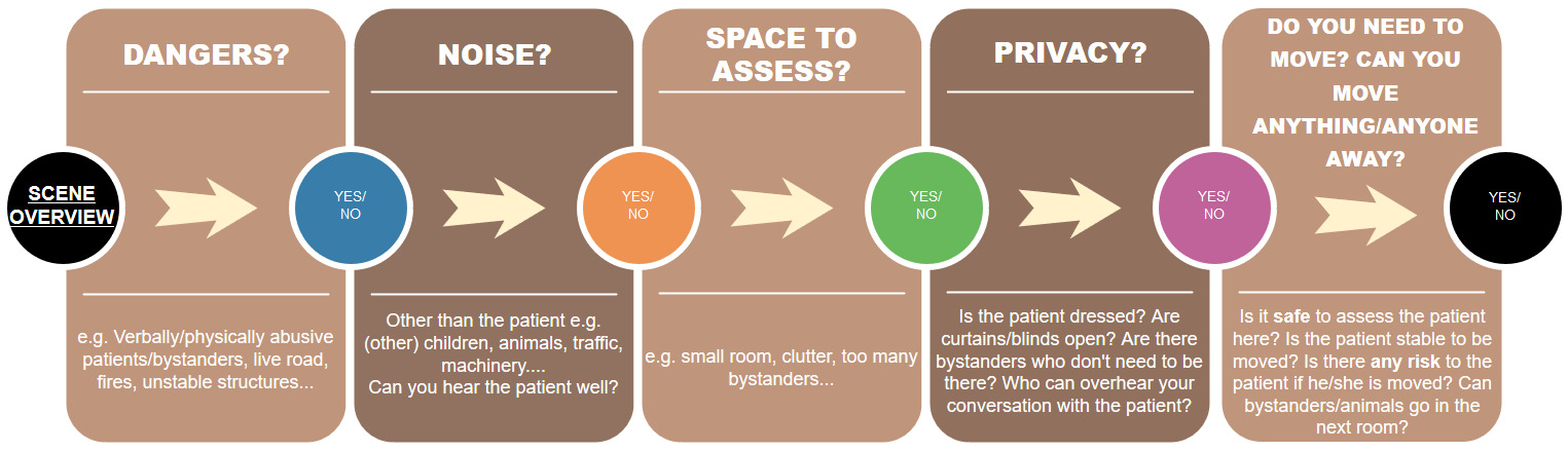
You may find varying suggestions for what to consider in what is usually referred to as your ‘scene assessment’ (Harris, 2016: 1; Pilbery & Lethbridge, 2016: 126) or ‘end of bed […] assessment’ (Spurr, 2014). This is a cheat sheet to assist you in identifying these important factors. There are a few examples within each category, forming part of what is surely an endless list. Harris (2016: 1-3) also recommends considering use of PPE, considering vehicle position, and considering CBRNE incidents. Pilbery & Lethbridge (2016: 127) recommend considering outside/inside temperature, terrain that is not reachable by land ambulance, and referring to major incident guides where applicable. Major incidents are not covered within this cheat sheet, but this sheet may guide you through the initial thought process prior to announcing a major incident…
Final Note & References
This is a cheat sheet which is designed to complement your scene assessment. This cheat sheet is in the author’s own words but ideas have been used from the below texts. Any copies of the above cheat sheets should be paired with the below references.
Harris, G. 2016. ‘General principles of assessment’ in A.Y. Blaber and G. Harris, ed. 2016. Assessment Skills for Paramedics, 2nd ed. Berkshire: Open University Press, pp. 1-13
Pilbery, R. and Lethbridge, K. 2016. Ambulance Care Practice, Bridgwater: Class Professional Publishing
Spurr, J. 2014. End-of-Bedogram: The Art Formally Known as Intuition, Available Online: http://injectableorange.com/2014/03/end-bedogram-art-formally-known-intuition/ (Accessed 26/03/18)
Feedback? Comments? Please post below…
This post is yet to be peer reviewed. Please get in touch if you have any comments.
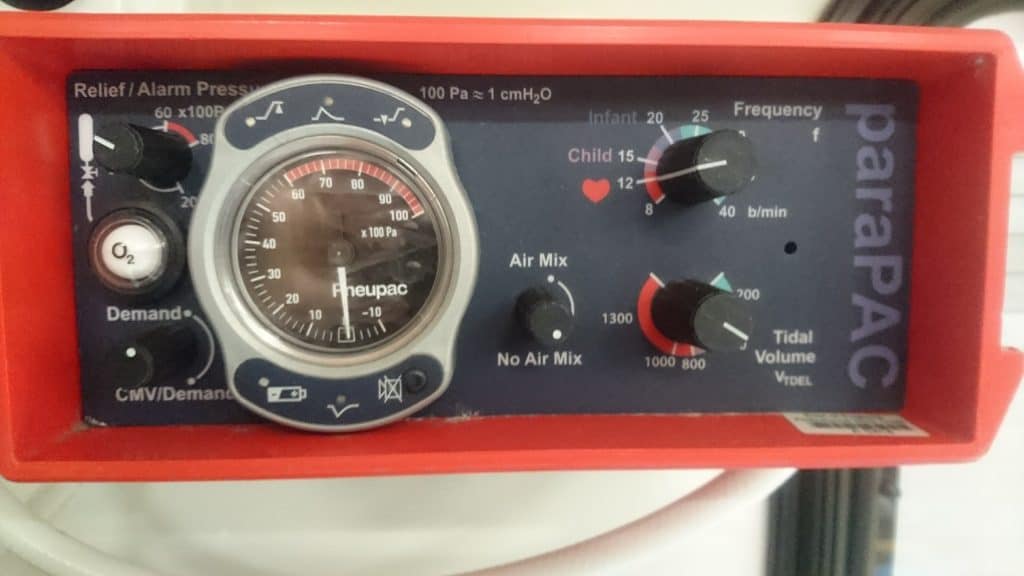
So far, Article 999 has a reasonable collection of information and videos about positive pressure breathing and ventilators used in prehospital emergency care. Smiths Medical is behind the PneuPac ParaPac. They have kindly given their permission to share the following documents, which are available for those who want to read more, find out about other available ventilators, or find an answer not available here.
A shorter version of Ventilators: Why, What, How, When? featuring how-to use the ventilator demonstrations only. Made by UK Paramedics, this demonstrates how to use the PneuPac ParaPac ventilator.
You must read the full disclaimer at www.article999.co.uk/about/ (disclaimer tab) before putting into place anything you see here. Useful information is available in the text on this video, so if you only listen to the narration you will miss important facts. The ventilation settings mentioned in this video are what is recommended by Smiths Medical, current guidelines and some articles – however, as with all topics, there is always varying information available online & alternative expert advice, and no video can cater for all of that. Similarly, this video is intended to demonstrate how to use equipment & to introduce or remind you to the Why, What and When of ventilators – not to tell you that you should or shouldn’t be using it. That is dependent on local guidelines, your research & your choice as a clinician. This video merely highlights the varying advice regarding tidal volume settings, and in the text points out the potential problems with some of the figures. No specific volume is recommended.
Transcript
Article 999. Ventilators: Why, What, How, When?
This video has been made by UK Paramedics following guidelines. It is not endorsed by any author, organisation or Ambulance Trust. You must read the full disclaimer at www.article999.co.uk/about/ and refer to your local guidelines before putting into place anything you see here. This video is intended to demonstrate how to use the ventilator according to use guides and guidelines – not to tell you that you should or shouldn’t use it. That decision is up to you and should be dependent on local guidelines and your own research. This is what the textbooks, the manufacturer and a few articles say & is not intended to represent the expert opinions or experience of others within healthcare.
Remember hand hygiene, bare below the elbows and gloves in real life.
How?
To remove the ventilator from the racks, pull the flap to the side and push the ventilator up. To put it back in, simply push it down and pull the flap to the side again.
To connect to the gas supply, push the hose into the socket and twist. It’s the same when connecting to a portable oxygen cylinder.
When you turn it on, the ventilator should complete a self check. The alarm lights should flash in sequence, there should be one burst of the high pressure audible alarm, and the orange indicator should flash for 60 seconds.
Next, check the indicator shows white for O2.
Connect the patient circuit [shown in video].
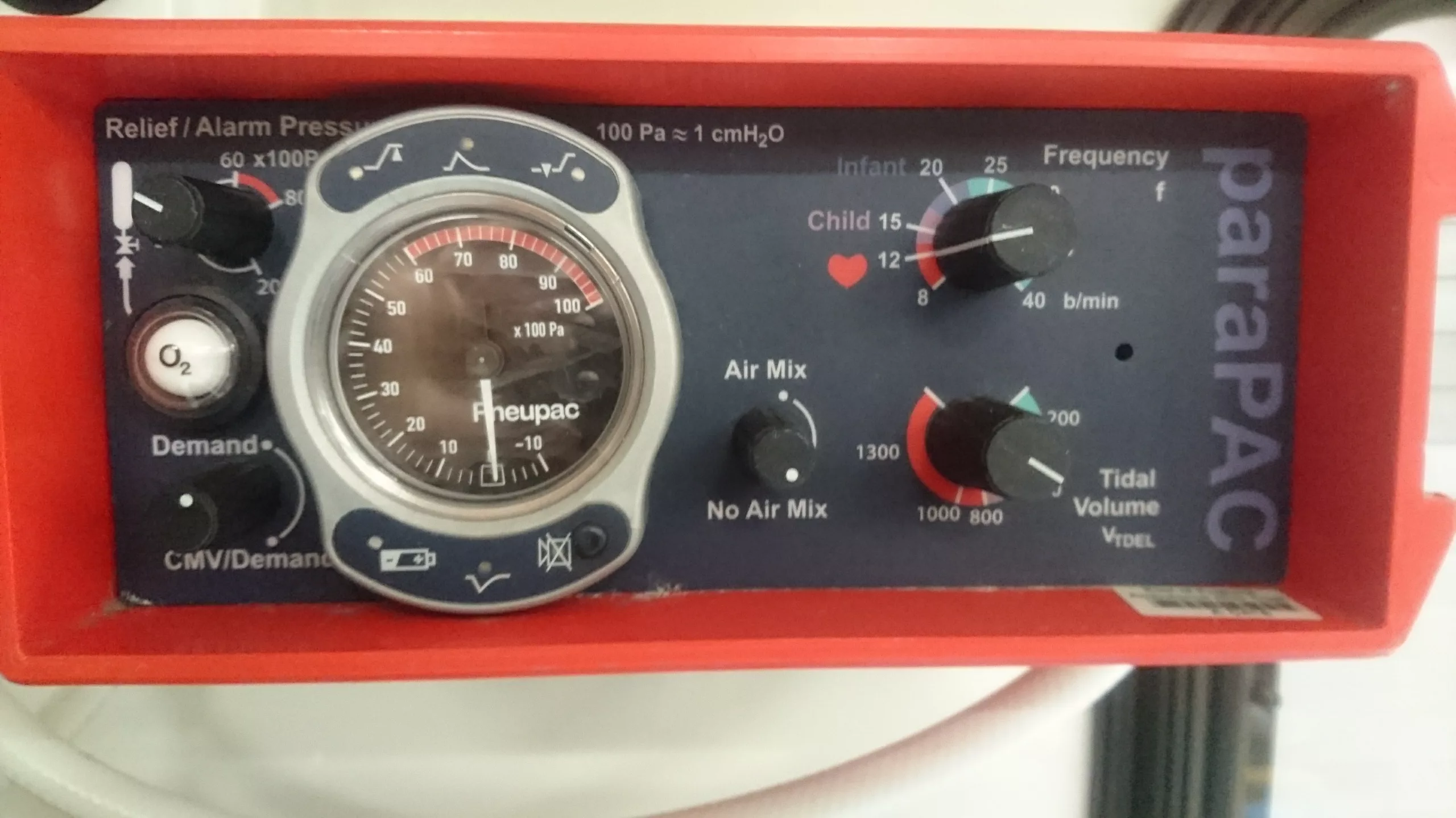
Next on the list, adjust the ventilation parameters. Set the air mix control to no air mix for respiratory arrest or CPR. Set the relief pressure control to 40. Set the respiratory rate to 12. As for the tidal volume, it varies massively as you can see on the screen. It depends on whether you opt for an average value or base it on the patient’s weight. Smiths Medical (2017) advise 800-900ml on their training video, [but this may not be advisable] but don’t worry too much about the slight differences in tidal volume because the PneuPac features a high pressure alarm that we’ll discuss shortly. The alarm will help you identify if the tidal volume is too high for your patient.
The middle one tells you when breathing is detected by the ventilator
(Smiths Medical, 2017)
High pressure = excessive tidal volume, incorrect airway position, kinked ET tube, or incorrect ventilation settings.
If the pressure is reaching it’s max, there may be something wrong with the circuit.
If you’ve checked the above and the alarm is still sounding, Smiths Medical (2017) advise that your tidal volume setting might simply be too high.
Low pressure = leakage or insufficient tidal volume/settings, faulty valve in the patient’s circuit
This alarm tends to occur when pressure drops below 10cmh20
Earlier, I pointed out the alarm in the middle, at the bottom. This is SMMV. It’s an indicator that will flash green if the patient is breathing for themselves. The ventilator will assist if the patient breathes with less than 150ml of tidal volume. Between 150-400ml the ventilator will extent the exhalation time to allow the patient to complete their own breath, and above 400ml the ventilator will allow the patient to breathe and will not assist, but may still assist on the next breath if required (Smiths Medical, 2017; Baker, 2012).
Extra facts:
McCarty et al (2012) found that ‘ventilation rates and tidal volumes commonly exceeded Guideline recommendations. This resulted in […] excessive mean airway pressure’
Smiths Medical (2017) advise that peak inflation pressure should be below 20cmh20 when using a mask to ventilate due to the issues of too high pressure
Inspiration to Expiration should be 1:2 (Smiths Medical, 2017; Baker, 2012)
References
Baker, D. 2012. Emergency and Transport Ventilation, an introductory guide, Smiths Medical International Limited: Luton
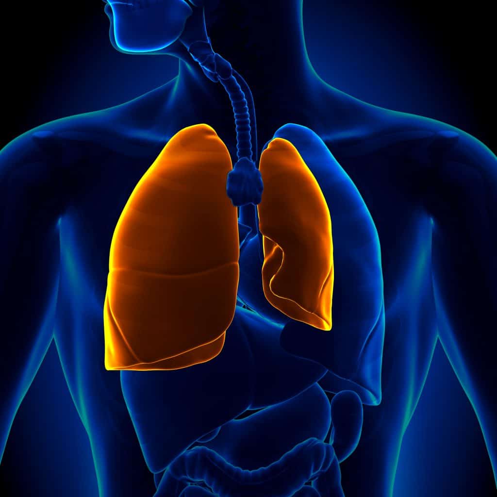
Positive pressure refers to the way artificial ventilation ‘inflate[s] the lungs’ (Hess & Kacmarek, 2014). ‘Normal breathing depends on drawing in air to the lungs by creating a partial vacuum inside the chest cavity’ (Baker, 2016: vi). The mean airway pressure, an average combined of ‘the amount and duration of pressure applied during’ expiratory and inspiratory phases in normal breathing is negative (Hess & Kacmarek, 2014: 420 ebook). See the article on negative pressure breathing for more information. Intermittent positive pressure ventilation, on the other hand, is for inspiration the ‘exact reverse of this process with air being forced into the lungs’ (Baker, 2016: vi).
Positive pressure ventilation includes the use of bag-valve-masks, but the majority of this article will focus on the ventilator.
Upcoming articles will discuss the specific settings and how-tos relating to the Pneupac ParaPac ventilator. Current links:
Ventilators have 4 phases of operation:
1. ‘The inspiratory phase’
2. ‘Cycling between the inspiratory (I) and expiratory (E) phase’
3. ‘The expiratory phase’
4. ‘Cycling between the E and I phase’
(Baker, 2016: 117)
The majority of ventilators are set to an I (Inspiratory) to E (Expiratory) ratio of 1:2, i.e. the expiratory phase is twice as long as the inspiratory phase, ‘which mirrors normal breathing’ (Baker, 2016: 108). If required, some ventilators allow this setting to ‘be altered to assist with restrictive lung diseases’ (Baker, 2016: 108). ICU ventilators contain additional settings that alter this ratio, including a ‘respiratory pause’ between the I and E phases ‘to allow distribution of gas within the lungs’ (Baker, 2016: 108).
[tabby title=”Mechanics”]
Mechanics
So, inspiration occurs when air is ‘forced into the lungs’ (Baker, 2016: vi). Expiration, however, occurs when ‘the ventilator stops delivering a positive pressure and the patient breaths out passively, as in normal breathing. This passive expiration (E) is ensured by the elastic recoil of the lungs and the chest wall’ (Baker, 2016: 108). The timing is aided by a ‘pneumatic oscillator’ (Baker, 2016: 22).
The gas is delivered by the ‘patient circuit’, which also ‘evacuate[s] expired CO2’ (Baker, 2016: 109). This is delivered ‘as close as possible to the airway device being used’ by a valve (Baker, 2016: 109). There are also filters to prevent contamination of the ventilator from the patient.
Early ventilators had a ‘lack of interactivity with the patient’s own breathing efforts if there was only partial respiratory failure’ (Baker, 2016: 22) but modern ventilators tend to have different modes to account for this and additional settings beyond a simple tidal volume dial. In particular, ventilators feature a ‘demand valve’ which detects when a patient takes a breath (Baker, 2016: 22). There are also settings for tidal volume and ventilation frequency. These settings will be discussed in upcoming articles.
The main positive effect of artificial ventilation is fairly obvious: it assists with the ventilation of a patient who is completely or partially unable to breathe normally. There are however a number of negative effects created by positive pressure ventilation. Related to these is the fact that mechanical ventilation can cause ‘heterogeneity’ of the lungs, meaning that ‘some lung units are prone to overdistension and others are prone to collapse’ (Hess & Kacmarek, 2014: 493). These are part of the reason most ventilators have relief pressure settings. This setting will be discussed in an upcoming article. You can check back here for the link.
Positive pressure ventilation can cause or effect the following:
The following are unavoidable results of ventilator use:
Decreased cardiac output
…Leading to ‘hypotension and potential tissue hypoxia. This effect is greatest with high mean airway pressure, high lung compliance, and low circulating blood volume’ (Hess & Kacmarek, 2014: 513). The high pressure ‘decreases venous return and right heart filling, which may reduce cardiac output’ (Hess & Kacmarek, 2014: 513).
This pressure is normally lowest ‘during inhalation’, so venous return would be highest. But with positive pressure ventilation, ‘venous return is greatest during exhalation’ as opposed to inhalation (Hess & Kacmarek, 2014: 513-520).
Renal and gastric effects
Mechanical ventilation reduces urine output due to ‘decreased renal perfusion’ which results from ‘decreased cardiac output’ (Hess & Kacmarek, 2014: 527). Unfortunately this can cause many of the issues common with fluid retention. Similarly, gastric distension can result, along with gastrointestinal bleeding’ (Hess & Kacmarek, 2014: 533).
Increased intracranial pressure
…in head injury patients, due to ‘a decrease in venous return, which increases intracranial blood volume and pressure’ (Hess & Kacmarek, 2014: 540).
Pneumonia may occur, especially due to invasive ventilation which is the form in emergency practice (non-invasive refers to ventilatory support such as CPAP). It ‘results from aspiration of oropharnygeal secretions around the cuff of the endotracheal tube’ (Hess & Kacmarek, 2014: 499).
The following can occur as a result of ventilator settings:
Hyperventilation and hypoventilation
These risk effecting the pH, and causing respiratory alkalosis (Hess & Kacmarek, 2014: 499)
Oxygen Toxicity due to high oxygen levels (Hess, 2014: 506)
A Shunt is usually partly decreased but settings can change this:
A shunt is ‘perfusion (blood flow) without ventilation’, which happens ‘when blood flows from the right heart to the left heart without participating in gas exchange’ (Hess & Kacmarek, 2014: 427). There are two different types of shunts: capillary and anatomic. The first occurs when the alveoli are not ventilated. One example of this is pneumonia (Hess & Kacmarek, 2014: 427). The latter type of shunt occurs when blood flow ‘completely bypasses the lungs’, which can sometimes happen with ‘congenital cardiac defects’ (Hess & Kacmarek, 2014: 427).
Positive Pressure Ventilation’s typical effect is to decrease capillary shunt by improving ‘the distribution of ventilation, particularly by improving the ventilation of previously underventilated areas of the lungs’ (Hess & Kacmarek, 2014: 427-446)
Despite the above improvements, if the settings are not ideal hypoxemia can result. This is the same outcome as a shunt but would in this case be caused by ‘redistribution of pulmonary blood flow to unventilated regions’ due to ‘overdistension of some lung units’ (Hess & Kacmarek, 2014: 324). Positive pressure ventilation can also increase anatomic shunt (Hess & Kacmarek, 2014: 441)
Also due to incorrect settings, the following can occur as a lack of pressure or volume:
Atelectasis occurs when the pressure or volume during or after artificial ventilation isn’t enough to inflate the lungs –> complete or partial collapse.
Atelectasis ‘is larger with obese patients and when a higher fraction of inspired oxygen (FI02) is used […]. During mechanical ventilation […] atelectasis may occur when lungs are underinflated due to low tidal volumes, or when compression occurs (such as patient position or obesity)’ (Open Anesthesia, 2017). If the volume or pressure isn’t enough to ‘prevent the alveoli from collapsing’ (Open Anesthesia, 2017) then regardless of ventilator type (pressure vs volume controlled) atelectasis may result.
The following can occur as a result of apparatus setup:
Dead Space can be increased by mechanical ventilation (Hess & Kacmarek, 2014: 471). It may be ‘reduced by an artificial airway (ETT) but is increased by increasing distance from the end of the ETT to the means of evacuating CO2 in mechanical ventilation’ (Baker, 2016: 111). Size, age, upright postures and neck extension also increase it (Baker, 2016: 111).
The following can occur due to overdistension:
Barotrauma = ‘alveolar rupture due to overdistension’, which can cause numerous problems including pneumothorax (Hess, 2014: 480). Volume generators have a ‘safety valve which activates at a preset pressure’ to avoid barotrauma’ (Baker, 2016: 118). This will be discussed in an upcoming article.
Ventilator-Induced lung injury caused by overdistension. Massively effected by ventilator settings for tidal volume and relief pressure (Hess, 2014: 493)
There are additional risks of positive pressure ventilation, particularly mechanical ventilation. Some of these are related to the use of airway adjuncts. Others are related to the removal of the ventilator. To read more, check out the references below.
[tabby title=”Types of Positive Pressure Ventilation”]
Types of Positive Pressure Ventilation
‘A spectrum of modes exist between total artificial ventilation and a patient who is still breathing spontaneously but with reduced efforts’ (Baker, 2016: 105).
There are two main types of ventilators:
– Pressure generated
– Volume generated
Pressure generators ‘provide a constant pressure during the inspiratory phase’ (Baker, 2016: 118). Volume generators release gas once a set volume has been reached. In each one, only the pressure or the volume is a constant. Everything else is relative and will adjust until the preset parameter of volume or pressure has been reached (Baker, 2016; Hess, 2014).
Issues
‘The problem of pressure generation is that it cannot guarantee an adequate Vt [volume] in situations where the lung mechanics are abnormal or where a guaranteed Vt is required due to life-threatening hypoxia. They are therefore not suitable for most emergency ventilation, with the notable exception of neonatal and infant ventilation where flow generators are contraindicated due to the fragility of the lung tissue’ (Baker, 2016: 118).
Additional Types
Most ventilators switch from inspiration to expiration by ‘time cycling’ (Baker, 2016: 119). However, this can also be due to volume or pressure cycling. The time cycling ratio is usually 1:2 for I:E, in other words the expiration time is twice as long as the inhalation time. ‘Ventilation modes can best be understood as a spectrum describing the work of breathing done by the ventilator and by the patient’ (Baker, 2016: 122.
The modes are: Controlled Mandatory Ventilation, in which ‘ventilation is provided entirely by the ventilator’ (Baker, 2016: 122), Assisted Controlled Ventilation which is fairly self-explanatory, and Synchronised Mandatory Ventilation, in which ‘the patient’s respiratory efforts in initiating a ventilation are synchronised with ventilations provided automatically by the ventilator.’ This is best used to supplement the patient’s efforts. It’s otherwise known as ‘assist-control’ and is typically found in ICU. (Baker, 2016: 123).
In portable ventilators the modes are:
Intermittent Mandatory Ventilation, Synchronised intermittent mandatory ventilation, and synchronised mandatory minute volume but only the latter two are still in use. The second overcomes the problems of the first, ensuring that breaths are supplemented by the ventilator and that supplemented breaths are activated only when there is a certain trigger e.g. low pressure. There is a modern, more complex version of this now in use in hospitals but this may exist in emergency ventilation practice to assist a patient who is ‘in partial respiratory failure’ (Baker, 2016: 124).
The first two divide minute volume between the ventilator and the second. The latter is based on time, which is compared with a ‘set minute volume. If there is a discrepancy between the actual minute volume and the set delivered minute volume, an automatic ventilation is delivered which corrects the difference’ (Baker, 2016: 124).
Because there is an issue of the ventilator being unable to tell the difference between dead space ventilation and alveolar ventilation, Pneupac have modified the above with a series of Ifs (if the patient takes a breath, if there is no further breath, if the tidal volume is too low, etc) that adjusts settings accordingly (Baker, 2016: 124). There are more modes that exist for ICU ventilators, which tend to be a lot more complex in design (Baker, 2016).
CPAP is also a form of positive pressure breathing and is designed ‘to improve oxygenation in lungs that may not be exchanging oxygen with the blood adequately’ (Baker, 2016: 126). There is a constant pressure in CPAP, versus ‘different pressures during inspiration and expiration’ in BIPAP’ but both are designed to allow the patient to do all the work of breathing. They merely increase oxygen and make inspiration easier while increasing the work of expiration. ‘Breathing with CPAP has been compared to the sensation of breathing against a very strong wind’ (Baker, 2016: 128). They ‘open up (recruit) alveoli in the lungs that may be non-ventilated.’ (Baker, 2016: 127). Both push back fluid ‘into the circulation via lymphatic drainage of the lung interstitial space’ and increase ‘the diffusion of oxygen into the pulmonary capillaries’ (Baker, 2016: 127).
[tabby title=”PEEP”]
PEEP
Positive End Expiratory Ventilation applies ‘a positive pressure to the patient during the expiratory phase’ to ‘increase the functional residual capacity of the lungs and thus the efficiency of oxygenation of the blood’ (Baker, 2016: 126). ‘In order to be able to deliver a set tidal volume against variable lung compliance and airway resistance, resuscitation ventilators should be time-cycled, volume preset flow generators’ (Baker, 2016: 132).