Category: I am a Medic

Content for paramedics, student paramedics, apprentice EMTs, and ECAs

  • Methods of ECG Interpretation

    Methods of ECG Interpretation

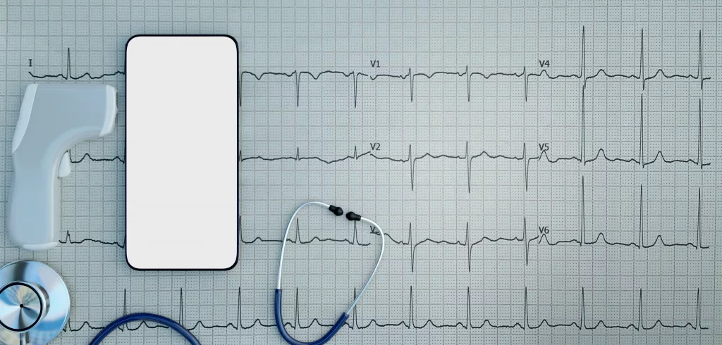
    There are so many different recommendations of ECG Interpretation. Some books have 5 steps, some 6, some 9, some 11. Below, I have listed a few common ones in the hope of highlighting the differences – and the similarities. I have tried to focus on rhythm strip analysis only but some methods combine the rhythm strip with the 12 lead analysis. I have only included those that are publicly available, so although some fantastic methods are taught on courses, I can’t reference them and have subsequently omitted them. However, if you have a preference or believe one method should be included below, add it in the comments and it may be featured in a future update.
    Method one:

    From: Resuscitation Council, Advanced Life Support (2021: 106):

    The 6 Steps of rhythm strip analysis:

    1. Is there any electrical activity? (Or/and check a pulse)

    2. What is the ventricular (QRS) rate?

    3. Is the QRS rhythm regular or irregular?

    4. Is the QRS complex width normal (‘narrow’) or broad?


    5. Is atrial activity present?

    6. Is atrial activity related to ventricular activity and, if so, how?

    Method Two:

    From: Charles L. Till, Clinical ECGs in Paramedic Practice (2021: 16):

    9 Steps – initially look at lead II, then look at every ECG lead:


    ECG Steps
    1. What is the rate and rhythm?
    2. Are there any P waves and what is their relationship with the QRS complex?
    3. What is the duration and morphology of the QRS complex?

    4. Is the ST segment isoelectric, depressed or elevated?
    5. Are the QT intervals and T waves normal?
     

    Clinical Steps
    6. Is the heart generating a palpable pulse of appropriate rate and providing adequate perfusion?
    7. Is the rhythm unstable and at risk of deterioration?
    8. Does the presenting rhythm support or change your working diagnosis?
    9. Are any clinical interventions required?
    Method Three:

    From: Paul Murray, East of England Ambulance Service, Version 1.0, ECG Recognition – Quick Reference Guide (Feb 2014: 1)

    6 steps:

    1. ‘QRS Rate’

    2. ‘Is the QRS rhythm regular or irregular?’ (and regularly irregular or irregularly irregular?)

    3. ‘QRS duration’

    4. ‘Are P waves present?’

    5. ‘Relationship between P waves and QRS – is there a P wave for each QRS and a QRS for each P wave?’

    6. ‘Is the PR interval within normal limits 120-200ms and is it constant?’

    (This is basically the same as the RC council method, with one added step; the RC method has one step at the start – a reminder to ensure the leads are connected properly and the patient is alive).
    Method Four

    From: Fred Kusumoto & Pam Bernath, ECG Interpretation for Everyone – An On-The-Spot Guide (2012: 33-36):

    ‘Assess the patient: symptoms and physical examination. Appearance, vital signs, physical examination’ ->
    1. Is there a P wave in front of every QRS and is the rate between 50-100 beats per minute?
    2. Are the ST segments isoelectric?

    Each answer leads you to a different figure – an option to confirm you have a normal ECG, evaluation of ST segments, evaluation of arrhythmias, or confirmation time-critical patients.

    The normal ECG should have:
    1. Rate should be between 50 and 100bpm

    2. A P before every QRS (Positive P in lead II).

    3. The QRS in V1 should be narrow an negative i.e. an Rs

    4. The ST segment should be isoelectric

    5. The T wave should be the same direction as the QRS
    Method Five:

    From: Kuhn, Lang, and Wiesbauer, ECG Mastery: The Simplest Way to Learn the ECG (2014: 141-142)

    Summarised – 11 steps (taking you from rhythm analysis into 12 lead analysis. Steps included to highlight how some of the steps above are readdressed or appear later altogether here; other points may appear in front):
    1. Rhythm. Criteria for sinus rhythm:

    • Are the P waves positive in I and II?
    • Is there a QRS complex after each P wave?
    • Are the PR intervals constant?
    • Are the RR intervals constant?

    Ask: Is it sinus?

    2. Heart rate

    3. P waves – atrial enlargement present?

    4. PR interval

    5. QRS axis

    6. QRS duration

    7. Rotation

    8. QRS amplitude

    9. QRS infarction signs

    10. ST-T segment

    11. QT duration, T-U waves


    That’s it for today, though you may find other methods at useful courses such as Mark Whitbread’s ECG Bootcamp.

    Do you have any recommendations, preferences, or opinions? Add them below!
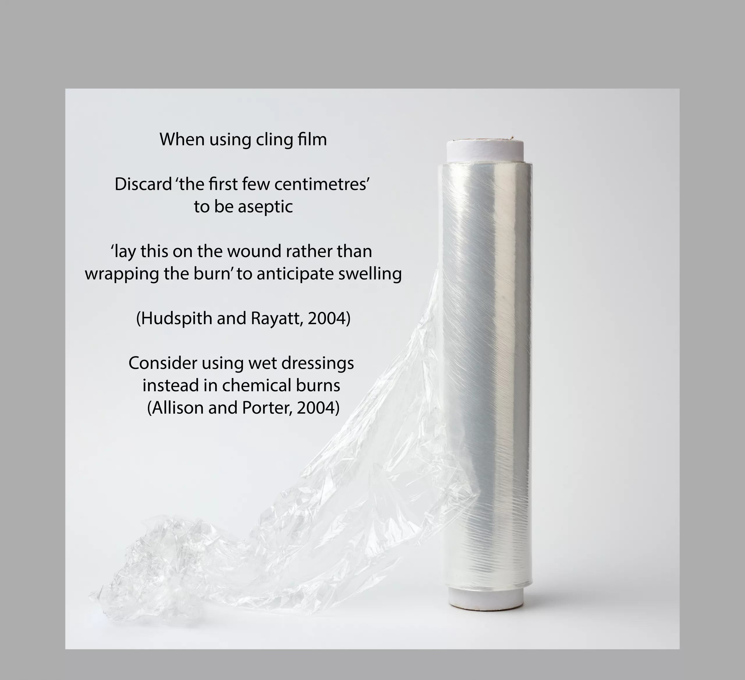
  • How to Use Cling Film to Treat Burns (Fast Fact)

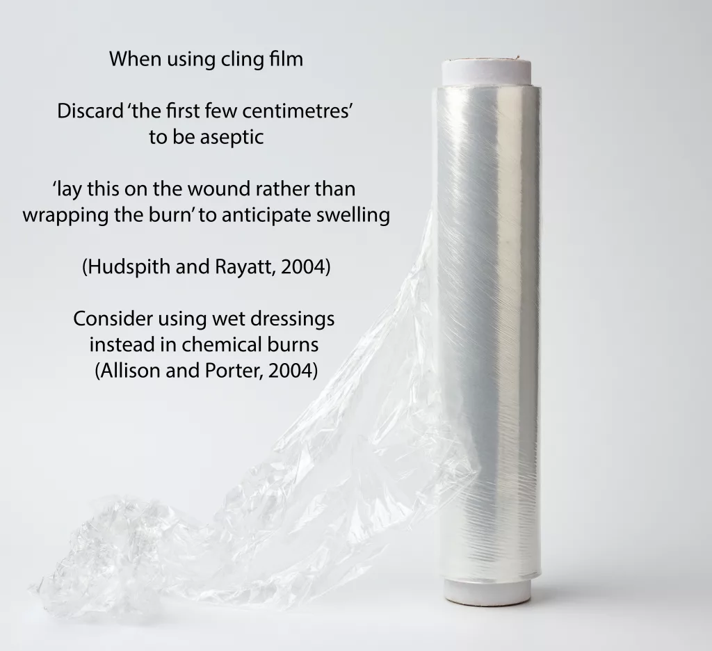
    How to Use Cling Film to Treat Burns (Fast Fact)

    Fast Fact posts are designed to provide quick and easy ways to revise key topics – view a photo with a quote/fact by simply scrolling through the category. Refresh yourself during those middle-of-the-night breaks when you would have aimlessly scrolled through your phone. There is no need to read a lengthy post to do a little CPD. For more fast facts, head here.

  • End of Bed Assessment – 16 Reasons Your Patient is Pale

    End of Bed Assessment – 16 Reasons Your Patient is Pale

    Here’s a list I’ve had for a while but not published: obvious, less obvious, and utterly surprising reasons your alive patient might be pale. References within. Detailed reference list coming soon.

    Image above – a person with vitiligo (Source: Adobe Stock)
    1. Shortness of breath (which may also be due to anaemia) – https://www.ncbi.nlm.nih.gov/pmc/articles/PMC2996160/
    2. Distributive, Cardiogenic, Obstructive + Dissociative shocks (Pilbery & Lethbridge, 2016: 247)
      • Including SOB/DIB, PE, pneumothorax, anaphylaxis
    3. Lung disease, leading to central cyanosis (Douglas et al, 2013: 45) including asthma and COPD (exacerbations & chronic)
    4. Acute myocardial infarction – https://patient.info/doctor/acute-myocardial-infarction
    5. Heart failure – https://em.osumc.edu/education/journalClub/SignsandSymptomsofHeartFailure.pdf
    6. Shock, caused by hypotension – https://www.nhlbi.nih.gov/health-topics/hypotension
    7. Vasovagal – ‘Facial pallor is often the first sign of an impending vasovagal faint’. https://academic.oup.com/brain/article/132/10/2630/329792>
    8. Shock – hypovolaemia – https://artifactsjournal.missouri.edu/2016/04/hypovolemic-shock-and-fluid-resuscitation/
    9. Lingual Raynaud Phenomenon – leading to a white tongue, temporarily – http://www.cmaj.ca/content/188/15/E396
    10. Drugs – Amiodarone can cause a ‘bluish-grey’ skin discoloration (Douglas et al, 2013: 44)
    11. Vitiligo (segmental and non-segmental) due to the lack of melanin, causes ‘pale patches of skin’ (Douglas et al, 2013: 46); non-segmental vitiligo ‘is thought to be an autoimmune condition’ (NHS – Vitiligo)
    12. Albinism (Douglas et al, 2013: 46)

    Discoloration of the hands/nails/eyelids:

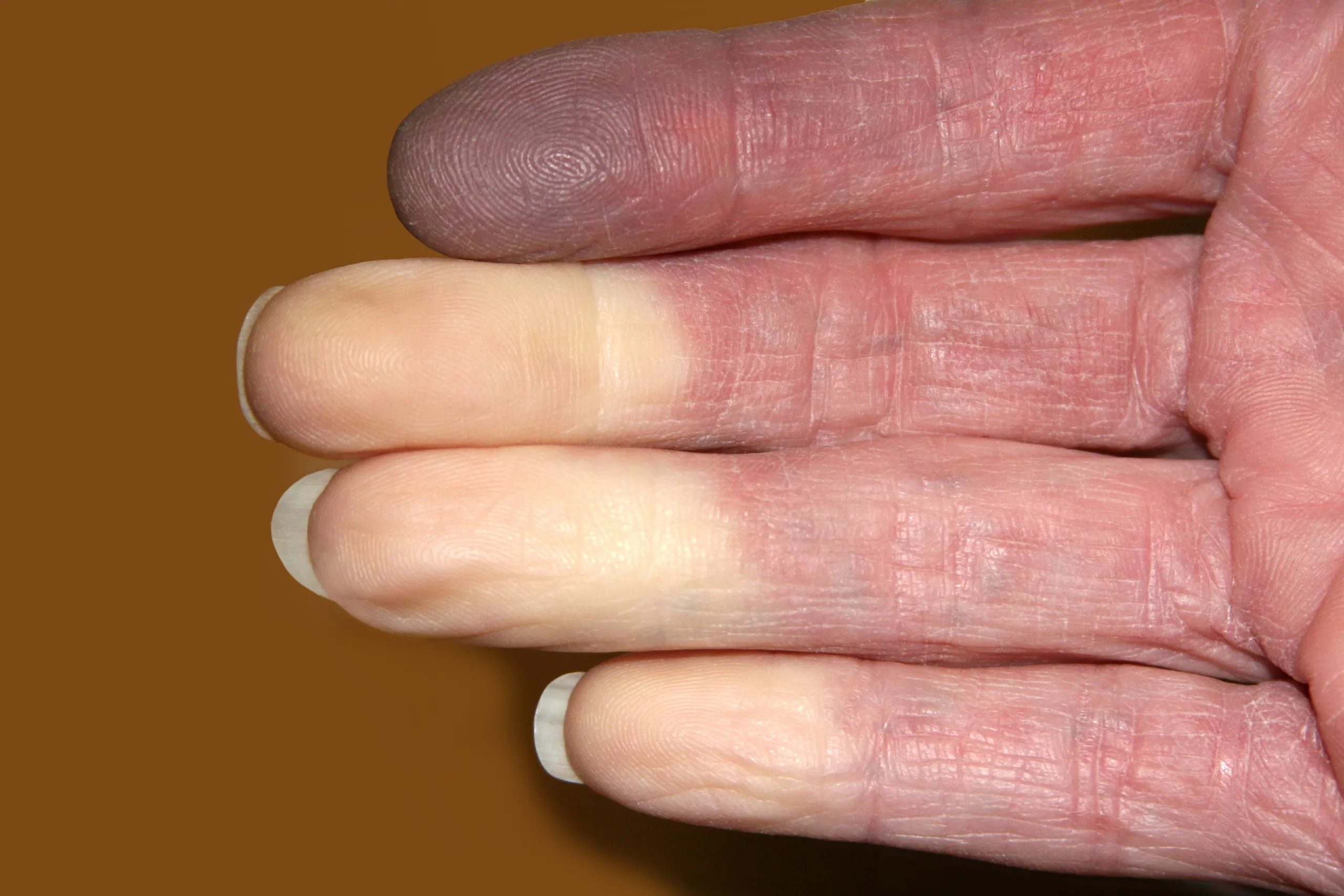
    Photo above demonstrates reynauds (Source: Adobe Stock)

    13. White discoloration of nails – 6 of 155 HIV patients in a 1998 study had this, amongst other more prevalent changes of their nails  – https://jamanetwork.com/journals/jamadermatology/fullarticle/189490

    14. Anaemia – ‘pallor of the conjunctiva, palm, nail beds or at any site was associated with a significantly lower hemoglobin concentration’ – From <https://academic.oup.com/jn/article/129/9/1675/4721973>

    15. Raynaud Phenomenon – leading to white/yellow/purple fingers, temporarily – http://www.cmaj.ca/content/188/15/E396

    Red herring

    16. Rigor mortis and liver mortis in an alive patient who was suffering from a dissecting abdominal aorta: https://content.sciendo.com/view/journals/sjfs/22/1/article-p11.xml

    Have I missed any? Add in the comments below —>

  • Acute Diarrhoea in the Elderly – The Risk Factors

    Acute Diarrhoea in the Elderly – The Risk Factors

    Sign, symptom, or conditionSign of, and TreatmentReference
    Sunken eyesDehydration; Patient may need referral to Doctor or urgent rehydration, depending on severity. Patient may benefit from blood testsJohnson and Smith, 2012: 94.
    High NEWS2 ScoreSepsis; patient may need antibiotics and emergency treatmentRoyal College of Physicians, 2017  
    Covid-19The diarrhoea could be a sign of deterioration, but the evidence for this is weakAmico et al, 2020  
    Female and increased ageIncreased risk of dehydrationRowat et al, cited in Sweetser, 2012
    Heart or kidney failure; take steroidsIncreased risk of overhydration; patient may require emergency treatmentSweetser, 2012; WHO, 2021.
    DizzinessDehydration; patient may require urgent rehydrationSweetser, 2012; WHO, 2021
    Confusion; seizuresDehydration or overhydration; electrolyte imbalances; patient may require emergency treatment and urgent rehydrationSweetser, 2012; WHO, 2021
    Type 2 Diabetes; metforminMetformin can cause the symptoms; diabetes is a risk factor due to potential difficulties managing the conditionJohnson and Smith, 2012: 96
    ImmunosuppressedRisk of deteriorationJohnson and Smith, 2012: 96
    Bowel disease such as ulcerative colitis or diverticulitisMay indicate more severe illnessJohnson and Smith, 2012: 96
    Blood in stoolsMay be an indication of abdominal bleedingJohnson and Smith, 2012: 96
  • ‘Thanks for the Feedback’ – A Video Summary in Quotes

    ‘Thanks for the Feedback’ – A Video Summary in Quotes

    For mentors/PPEDs, PHEM feedback facilitators, ALS instructors, teachers, and those interested in responding better to feedback (and in doing so, giving better feedback), here’s one for you. This is a summary in quotes of the ‘Thanks for the Feedback’ book, picking up on some key points. The full reference is available in our new reference database here: https://airtable.com/shrmbfb43bwcyYtPG

    This video took about 2 hours of an evening to produce and was created by Article 999’s founder, Louise, using a great software program purchased out of pocket. The actual reading and saving of quotes has taken [insert actual time] far too long and a similar amount of time will be needed for other books. If you like Article 999’s content, let us know by buying us a coffee here or clicking on the coffee symbol in the bottom right corner.

  • HCPC CPD Portfolio Help Folder and Template

    HCPC CPD Portfolio Help Folder and Template

    If you’re a UK Paramedic looking for a template to use for your CPD portfolio, here’s a link to one I’ve made. You can download and copy this template into your OneNote or other note application. Simply fill in the blanks, create your own dated list of CPD activities*, and then delete the help info. Keep your completed portfolio available for any HCPC audits, and save the help folder for any questions. The portfolio is referenced and contains direct links with screenshots from useful sources.

    *I now also have a free template available for you to use to add your own CPD. Please follow this link for more information. (Feb 2022; link updated Jan 2023)

    Let me know what you think.

  • What Does NICE Say About Epilepsy? The Video

    What Does NICE Say About Epilepsy? The Video

    The guidelines, summarised, and made relevant for Paramedics (anything irrelevant is not included):

    The full guidelines:

    https://www.nice.org.uk/guidance/cg137/resources/epilepsies-diagnosis-and-management-pdf-35109515407813

    (full reference in video)

    *update 28/02/22 – music volume reduced to ensure voiceover is clearer